 Movie
Movie Controller

[English] 日本語
 Yorodumi
- PDB-6s67: Structure of the Fluorescent Protein AausFP1 from Aequorea cf. au... -
+ Open data

- Basic information
| Entry | Database: PDB / ID: 6s67 | ||||||
|---|---|---|---|---|---|---|---|
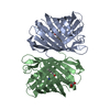
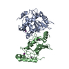
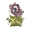
| Title | Structure of the Fluorescent Protein AausFP1 from Aequorea cf. australis at pH 7.0 | ||||||
 Components Components | Aequorea cf. australis fluorescent protein 1 (AausFP1) | ||||||
 Keywords Keywords |  FLUORESCENT PROTEIN / GFP-like fluorescent protein FLUORESCENT PROTEIN / GFP-like fluorescent protein | ||||||
| Function / homology | Green fluorescent protein, GFP / Green fluorescent protein-related / Green fluorescent protein / Green fluorescent protein / bioluminescence / generation of precursor metabolites and energy / Green fluorescent protein FP1 Function and homology information Function and homology information | ||||||
| Biological species |  Aequorea australis (invertebrata) Aequorea australis (invertebrata) | ||||||
| Method | X-RAY DIFFRACTION / SYNCHROTRON / MOLECULAR REPLACEMENT / molecular replacement / Resolution: 2.47 Å | ||||||
 Authors Authors | Depernet, H. / Gotthard, G. / Lambert, G.G. / Shaner, N.C. / Royant, A. | ||||||
| Funding support |  United States, 1items United States, 1items
| ||||||
 Citation Citation | Journal: Plos Biol. / Year: 2020 Title: Aequorea's secrets revealed: New fluorescent proteins with unique properties for bioimaging and biosensing. Authors: Lambert, G.G. / Depernet, H. / Gotthard, G. / Schultz, D.T. / Navizet, I. / Lambert, T. / Adams, S.R. / Torreblanca-Zanca, A. / Chu, M. / Bindels, D.S. / Levesque, V. / Nero Moffatt, J. / ...Authors: Lambert, G.G. / Depernet, H. / Gotthard, G. / Schultz, D.T. / Navizet, I. / Lambert, T. / Adams, S.R. / Torreblanca-Zanca, A. / Chu, M. / Bindels, D.S. / Levesque, V. / Nero Moffatt, J. / Salih, A. / Royant, A. / Shaner, N.C. | ||||||
| History |
|
- Structure visualization
| Structure viewer | Molecule: MolmilJmol/JSmol |
|---|
- Downloads & links
-Download
| PDBx/mmCIF format | 6s67.cif.gz | 197.3 KB | Display | PDBx/mmCIF format |
|---|---|---|---|---|
| PDB format | pdb6s67.ent.gz | 156.7 KB | Display | PDB format |
| PDBx/mmJSON format | 6s67.json.gz | Tree view | PDBx/mmJSON format | |
| Others |  Other downloads Other downloads |
-Validation report
| Arichive directory | https://data.pdbj.org/pub/pdb/validation_reports/s6/6s67ftp://data.pdbj.org/pub/pdb/validation_reports/s6/6s67 | HTTPS FTP |
|---|
-Related structure data
| Related structure data |  6s68C 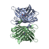 4he4S S: Starting model for refinement C: citing same article ( |
|---|---|
| Similar structure data |


-Links
 PDBj
PDBj

- Assembly
| Deposited unit | 
| |||||||||||||||||||||||||||||||||||||||||||||||||||||||||||||||||||||||||||||||||||||||||||||||||||||||||||||||||||||||||||||||||||||||||||||||||||||||||||||||||||||||||||||||||||||||||||||||||||||||||||||||||||||
|---|---|---|---|---|---|---|---|---|---|---|---|---|---|---|---|---|---|---|---|---|---|---|---|---|---|---|---|---|---|---|---|---|---|---|---|---|---|---|---|---|---|---|---|---|---|---|---|---|---|---|---|---|---|---|---|---|---|---|---|---|---|---|---|---|---|---|---|---|---|---|---|---|---|---|---|---|---|---|---|---|---|---|---|---|---|---|---|---|---|---|---|---|---|---|---|---|---|---|---|---|---|---|---|---|---|---|---|---|---|---|---|---|---|---|---|---|---|---|---|---|---|---|---|---|---|---|---|---|---|---|---|---|---|---|---|---|---|---|---|---|---|---|---|---|---|---|---|---|---|---|---|---|---|---|---|---|---|---|---|---|---|---|---|---|---|---|---|---|---|---|---|---|---|---|---|---|---|---|---|---|---|---|---|---|---|---|---|---|---|---|---|---|---|---|---|---|---|---|---|---|---|---|---|---|---|---|---|---|---|---|---|---|---|---|
| 1 | 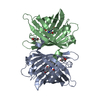
| |||||||||||||||||||||||||||||||||||||||||||||||||||||||||||||||||||||||||||||||||||||||||||||||||||||||||||||||||||||||||||||||||||||||||||||||||||||||||||||||||||||||||||||||||||||||||||||||||||||||||||||||||||||
| 2 | 
| |||||||||||||||||||||||||||||||||||||||||||||||||||||||||||||||||||||||||||||||||||||||||||||||||||||||||||||||||||||||||||||||||||||||||||||||||||||||||||||||||||||||||||||||||||||||||||||||||||||||||||||||||||||
| Unit cell |
| |||||||||||||||||||||||||||||||||||||||||||||||||||||||||||||||||||||||||||||||||||||||||||||||||||||||||||||||||||||||||||||||||||||||||||||||||||||||||||||||||||||||||||||||||||||||||||||||||||||||||||||||||||||
| Noncrystallographic symmetry (NCS) | NCS domain:
NCS domain segments: Component-ID: 0 / Refine code: 0
|