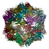
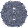
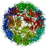
Journal: PLoS Pathog / Year: 2023 Title: Structure-guided mutagenesis of the capsid protein indicates that a nanovirus requires assembled viral particles for systemic infection. Authors: Stefano Trapani / Eijaz Ahmed Bhat / Michel Yvon / Joséphine Lai-Kee-Him / François Hoh / Marie-Stéphanie Vernerey / Elodie Pirolles / Mélia Bonnamy / Guy Schoehn / Jean-Louis Zeddam / ...Authors: Stefano Trapani / Eijaz Ahmed Bhat / Michel Yvon / Joséphine Lai-Kee-Him / François Hoh / Marie-Stéphanie Vernerey / Elodie Pirolles / Mélia Bonnamy / Guy Schoehn / Jean-Louis Zeddam / Stéphane Blanc / Patrick Bron / Abstract: Nanoviruses are plant multipartite viruses with a genome composed of six to eight circular single-stranded DNA segments. The distinct genome segments are encapsidated individually in icosahedral ...Nanoviruses are plant multipartite viruses with a genome composed of six to eight circular single-stranded DNA segments. The distinct genome segments are encapsidated individually in icosahedral particles that measure ≈18 nm in diameter. Recent studies on the model species Faba bean necrotic stunt virus (FBNSV) revealed that complete sets of genomic segments rarely occur in infected plant cells and that the function encoded by a given viral segment can complement the others across neighbouring cells, presumably by translocation of the gene products through unknown molecular processes. This allows the viral genome to replicate, assemble into viral particles and infect anew, even with the distinct genome segments scattered in different cells. Here, we question the form under which the FBNSV genetic material propagates long distance within the vasculature of host plants and, in particular, whether viral particle assembly is required. Using structure-guided mutagenesis based on a 3.2 Å resolution cryogenic-electron-microscopy reconstruction of the FBNSV particles, we demonstrate that specific site-directed mutations preventing capsid formation systematically suppress FBNSV long-distance movement, and thus systemic infection of host plants, despite positive detection of the mutated coat protein when the corresponding segment is agroinfiltrated into plant leaves. These results strongly suggest that the viral genome does not propagate within the plant vascular system under the form of uncoated DNA molecules or DNA:coat-protein complexes, but rather moves long distance as assembled viral particles.
Empty: NO / Enveloped: NO / Isolate: STRAIN / Type: VIRION
Virus shell
Name: capsid / Triangulation number (T number): 3
Buffer solution
pH: 7
Specimen
Embedding applied: NO / Shadowing applied: NO / Staining applied: NO / Vitrification applied: YES
Vitrification
Cryogen name: ETHANE
-
Electron microscopy imaging
Experimental equipment
Model: Tecnai Polara / Image courtesy: FEI Company
Microscopy
Model: FEI POLARA 300
Electron gun
Electron source: FIELD EMISSION GUN / Accelerating voltage: 300 kV / Illumination mode: FLOOD BEAM
Electron lens
Mode: BRIGHT FIELDBright-field microscopy
Image recording
Electron dose: 40 e/Å2 / Film or detector model: GATAN K2 SUMMIT (4k x 4k)
Image scans
Movie frames/image: 40
-
Processing
EM software
ID
Name
Version
Category
10
RELION
3
finalEulerassignment
12
RELION
3
3Dreconstruction
13
PHENIX
1.15.2
modelrefinement
14
Coot
0.9-pre
modelrefinement
CTF correction
Type: PHASE FLIPPING AND AMPLITUDE CORRECTION
Symmetry
Point symmetry: I (icosahedral)
3D reconstruction
Resolution: 3.19 Å / Resolution method: FSC 0.143 CUT-OFF / Num. of particles: 5156 / Algorithm: FOURIER SPACE / Num. of class averages: 1 / Symmetry type: POINT
Atomic model building
Protocol: AB INITIO MODEL / Space: REAL
+
About Yorodumi
-
News
-
Feb 9, 2022. New format data for meta-information of EMDB entries
New format data for meta-information of EMDB entries
Version 3 of the EMDB header file is now the official format.
The previous official version 1.9 will be removed from the archive.
In the structure databanks used in Yorodumi, some data are registered as the other names, "COVID-19 virus" and "2019-nCoV". Here are the details of the virus and the list of structure data.
Jan 31, 2019. EMDB accession codes are about to change! (news from PDBe EMDB page)
EMDB accession codes are about to change! (news from PDBe EMDB page)
The allocation of 4 digits for EMDB accession codes will soon come to an end. Whilst these codes will remain in use, new EMDB accession codes will include an additional digit and will expand incrementally as the available range of codes is exhausted. The current 4-digit format prefixed with “EMD-” (i.e. EMD-XXXX) will advance to a 5-digit format (i.e. EMD-XXXXX), and so on. It is currently estimated that the 4-digit codes will be depleted around Spring 2019, at which point the 5-digit format will come into force.
The EM Navigator/Yorodumi systems omit the EMD- prefix.
Related info.:Q: What is EMD? / ID/Accession-code notation in Yorodumi/EM Navigator
Yorodumi is a browser for structure data from EMDB, PDB, SASBDB, etc.
This page is also the successor to EM Navigator detail page, and also detail information page/front-end page for Omokage search.
The word "yorodu" (or yorozu) is an old Japanese word meaning "ten thousand". "mi" (miru) is to see.
Related info.:EMDB / PDB / SASBDB / Comparison of 3 databanks / Yorodumi Search / Aug 31, 2016. New EM Navigator & Yorodumi / Yorodumi Papers / Jmol/JSmol / Function and homology information / Changes in new EM Navigator and Yorodumi
 Movie
Movie Controller


 Open data
 Basic information
 Components
 Capsid
  Keywords
 Function and homology information
 Authors
 France, 1items
  Citation
 Structure visualization
 Downloads & links
 Other downloads
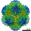


 PDBj
PDBj Assembly
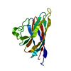

 Sample preparation
 Electron microscopy imaging
 Processing