 Movie
Movie Controller

[English] 日本語
 Yorodumi
- PDB-6s37: Ligand binding domain of the P. putida receptor PcaY_PP in comple... -
+ Open data

- Basic information
| Entry | Database: PDB / ID: 6s37 | |||||||||
|---|---|---|---|---|---|---|---|---|---|---|
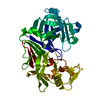
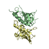
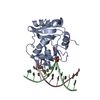
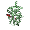
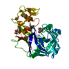
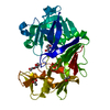
| Title | Ligand binding domain of the P. putida receptor PcaY_PP in complex with salicylic acid | |||||||||
 Components Components | Aromatic acid chemoreceptor | |||||||||
 Keywords Keywords |  SIGNALING PROTEIN / Ligand binding domain / Pseudomonas putida / chemotactic transducer / C6-ring carboxylic acids SIGNALING PROTEIN / Ligand binding domain / Pseudomonas putida / chemotactic transducer / C6-ring carboxylic acids | |||||||||
| Function / homology |  Function and homology informationchemotaxis / transmembrane signaling receptor activity / signal transduction / plasma membrane Function and homology informationchemotaxis / transmembrane signaling receptor activity / signal transduction / plasma membraneSimilarity search - Function | |||||||||
| Biological species |  Pseudomonas putida (bacteria) Pseudomonas putida (bacteria) | |||||||||
| Method | X-RAY DIFFRACTION / SYNCHROTRON / MOLECULAR REPLACEMENT / Resolution: 2.3 Å | |||||||||
 Authors Authors | Gavira, J.A. / Mantilla, M.A. / Fernandez, M. / Krell, T. | |||||||||
| Funding support |  Spain, 2items Spain, 2items
| |||||||||
 Citation Citation | Journal: Febs J. / Year: 2021 Title: The structural basis for signal promiscuity in a bacterial chemoreceptor. Authors: Gavira, J.A. / Matilla, M.A. / Fernandez, M. / Krell, T. | |||||||||
| History |
|
- Structure visualization
| Structure viewer | Molecule: MolmilJmol/JSmol |
|---|
- Downloads & links
-Download
| PDBx/mmCIF format | 6s37.cif.gz | 144 KB | Display | PDBx/mmCIF format |
|---|---|---|---|---|
| PDB format | pdb6s37.ent.gz | 107.4 KB | Display | PDB format |
| PDBx/mmJSON format | 6s37.json.gz | Tree view | PDBx/mmJSON format | |
| Others |  Other downloads Other downloads |
-Validation report
| Arichive directory | https://data.pdbj.org/pub/pdb/validation_reports/s3/6s37ftp://data.pdbj.org/pub/pdb/validation_reports/s3/6s37 | HTTPS FTP |
|---|
-Related structure data
| Related structure data |  6s18SC  6s1aC  6s33C  6s38C  6s3bC S: Starting model for refinement C: citing same article ( |
|---|---|
| Similar structure data |
-Links
 PDBj
PDBj


- Assembly
| Deposited unit | 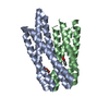
| ||||||||
|---|---|---|---|---|---|---|---|---|---|
| 1 |
| ||||||||
| Unit cell |
|
-Components
| #1: Protein | Mass: 59541.609 Da / Num. of mol.: 2 Source method: isolated from a genetically manipulated source Details: Ligand binding domain Source: (gene. exp.) Pseudomonas putida (strain ATCC 47054 / DSM 6125 / NCIMB 11950 / KT2440) (bacteria)Strain: ATCC 47054 / DSM 6125 / NCIMB 11950 / KT2440 / Gene: pcaY, PP_2643 Production host: Escherichia coli 'BL21-Gold(DE3)pLysS AG' (bacteria)References: UniProt: Q88JK6 #2: Chemical | ChemComp-SAL / | Salicylic acid  Mass: 138.121 Da / Num. of mol.: 1 / Source method: obtained synthetically / Formula: C7H6O3 / Feature type: SUBJECT OF INVESTIGATION Mass: 138.121 Da / Num. of mol.: 1 / Source method: obtained synthetically / Formula: C7H6O3 / Feature type: SUBJECT OF INVESTIGATION#3: Chemical | ChemComp-ACT / | Acetate  Mass: 59.044 Da / Num. of mol.: 1 / Source method: obtained synthetically / Formula: C2H3O2 Mass: 59.044 Da / Num. of mol.: 1 / Source method: obtained synthetically / Formula: C2H3O2#4: Water | ChemComp-HOH / | Water Mass: 18.015 Da / Num. of mol.: 54 / Source method: isolated from a natural source / Formula: H2O Mass: 18.015 Da / Num. of mol.: 54 / Source method: isolated from a natural source / Formula: H2OHas ligand of interest | Y | |
|---|
-Experimental details
-Experiment
| Experiment | Method: X-RAY DIFFRACTION / Number of used crystals: 1 |
|---|
- Sample preparation
| Crystal | Density Matthews: 1.8 Å3/Da / Density % sol: 33 % |
|---|---|
| Crystal grow | Temperature: 293 K / Method: counter-diffusion / pH: 6.5 Details: 30% PEG 8K, 0.1M Na Acetate, 0.1M Na-Cacodylate pH 6.50 |
-Data collection
| Diffraction | Mean temperature: 100 K / Serial crystal experiment: N |
|---|---|
| Diffraction source | Source: SYNCHROTRON / Site: ALBA / Beamline: XALOC / Wavelength: 0.979 Å |
| Detector | Type: DECTRIS PILATUS 6M / Detector: PIXEL / Date: Dec 5, 2018 |
| Radiation | Protocol: SINGLE WAVELENGTH / Monochromatic (M) / Laue (L): M / Scattering type: x-ray |
| Radiation wavelength | Wavelength: 0.979 Å / Relative weight: 1 |
| Reflection | Resolution: 2.3→40.44 Å / Num. obs: 13404 / % possible obs: 98.97 % / Redundancy: 5.3 % / Biso Wilson estimate: 36.33 Å2 / CC1/2: 0.998 / Rmerge(I) obs: 0.114 / Rpim(I) all: 0.054 / Rrim(I) all: 0.127 / Net I/σ(I): 12.44 |
| Reflection shell | Resolution: 2.3→2.382 Å / Redundancy: 5.1 % / Rmerge(I) obs: 1.14 / Mean I/σ(I) obs: 1.58 / Num. unique obs: 1321 / CC1/2: 0.627 / Rpim(I) all: 0.559 / Rrim(I) all: 1.27 / % possible all: 99.47 |
- Processing
| Software |
| |||||||||||||||||||||||||||||||||||||||||||||||||||||||||||||||||||||||||||||||||||||||||||||||||||||||||||||||||||||||||||||||||||||||||||||||||||||||||||||||||||||||||||||||
|---|---|---|---|---|---|---|---|---|---|---|---|---|---|---|---|---|---|---|---|---|---|---|---|---|---|---|---|---|---|---|---|---|---|---|---|---|---|---|---|---|---|---|---|---|---|---|---|---|---|---|---|---|---|---|---|---|---|---|---|---|---|---|---|---|---|---|---|---|---|---|---|---|---|---|---|---|---|---|---|---|---|---|---|---|---|---|---|---|---|---|---|---|---|---|---|---|---|---|---|---|---|---|---|---|---|---|---|---|---|---|---|---|---|---|---|---|---|---|---|---|---|---|---|---|---|---|---|---|---|---|---|---|---|---|---|---|---|---|---|---|---|---|---|---|---|---|---|---|---|---|---|---|---|---|---|---|---|---|---|---|---|---|---|---|---|---|---|---|---|---|---|---|---|---|---|---|
| Refinement | Method to determine structure: MOLECULAR REPLACEMENT Starting model: 6S18 Resolution: 2.3→40.44 Å / SU ML: 0.27 / Cross valid method: FREE R-VALUE / σ(F): 1.34 / Phase error: 23.64
| |||||||||||||||||||||||||||||||||||||||||||||||||||||||||||||||||||||||||||||||||||||||||||||||||||||||||||||||||||||||||||||||||||||||||||||||||||||||||||||||||||||||||||||||
| Solvent computation | Shrinkage radii: 0.9 Å / VDW probe radii: 1.11 Å | |||||||||||||||||||||||||||||||||||||||||||||||||||||||||||||||||||||||||||||||||||||||||||||||||||||||||||||||||||||||||||||||||||||||||||||||||||||||||||||||||||||||||||||||
| Refinement step | Cycle: LAST / Resolution: 2.3→40.44 Å
| |||||||||||||||||||||||||||||||||||||||||||||||||||||||||||||||||||||||||||||||||||||||||||||||||||||||||||||||||||||||||||||||||||||||||||||||||||||||||||||||||||||||||||||||
| Refine LS restraints |
| |||||||||||||||||||||||||||||||||||||||||||||||||||||||||||||||||||||||||||||||||||||||||||||||||||||||||||||||||||||||||||||||||||||||||||||||||||||||||||||||||||||||||||||||
| LS refinement shell |
| |||||||||||||||||||||||||||||||||||||||||||||||||||||||||||||||||||||||||||||||||||||||||||||||||||||||||||||||||||||||||||||||||||||||||||||||||||||||||||||||||||||||||||||||
| Refinement TLS params. | Method: refined / Refine-ID: X-RAY DIFFRACTION
| |||||||||||||||||||||||||||||||||||||||||||||||||||||||||||||||||||||||||||||||||||||||||||||||||||||||||||||||||||||||||||||||||||||||||||||||||||||||||||||||||||||||||||||||
| Refinement TLS group |
|
