Movie
Movie Controller


+ Open data

- Basic information
| Entry | Database: PDB / ID: 6s2o | ||||||
|---|---|---|---|---|---|---|---|
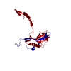
| Title | Granulovirus occlusion bodies by serial electron diffraction | ||||||
 Components Components | Granulin | ||||||
 Keywords Keywords | VIRAL PROTEIN / granulovirus / occlusion body / serial crystallography | ||||||
| Function / homology | Polyhedrin / Polyhedrin / viral occlusion body / structural molecule activity / Granulin Function and homology information Function and homology information | ||||||
| Biological species |  Cydia pomonella granulosis virus Cydia pomonella granulosis virus | ||||||
| Method | ELECTRON CRYSTALLOGRAPHY / electron crystallography / cryo EM / Resolution: 1.6 Å | ||||||
 Authors Authors | Buecker, R. / Mehrabi, P. / Schulz, E.C. / Hogan-Lamarre, P. | ||||||
 Citation Citation | Journal: Nat Commun / Year: 2020 Title: Serial protein crystallography in an electron microscope. Authors: Robert Bücker / Pascal Hogan-Lamarre / Pedram Mehrabi / Eike C Schulz / Lindsey A Bultema / Yaroslav Gevorkov / Wolfgang Brehm / Oleksandr Yefanov / Dominik Oberthür / Günther H Kassier / ...Authors: Robert Bücker / Pascal Hogan-Lamarre / Pedram Mehrabi / Eike C Schulz / Lindsey A Bultema / Yaroslav Gevorkov / Wolfgang Brehm / Oleksandr Yefanov / Dominik Oberthür / Günther H Kassier / R J Dwayne Miller /   Abstract: Serial X-ray crystallography at free-electron lasers allows to solve biomolecular structures from sub-micron-sized crystals. However, beam time at these facilities is scarce, and involved sample ...Serial X-ray crystallography at free-electron lasers allows to solve biomolecular structures from sub-micron-sized crystals. However, beam time at these facilities is scarce, and involved sample delivery techniques are required. On the other hand, rotation electron diffraction (MicroED) has shown great potential as an alternative means for protein nano-crystallography. Here, we present a method for serial electron diffraction of protein nanocrystals combining the benefits of both approaches. In a scanning transmission electron microscope, crystals randomly dispersed on a sample grid are automatically mapped, and a diffraction pattern at fixed orientation is recorded from each at a high acquisition rate. Dose fractionation ensures minimal radiation damage effects. We demonstrate the method by solving the structure of granulovirus occlusion bodies and lysozyme to resolutions of 1.55 Å and 1.80 Å, respectively. Our method promises to provide rapid structure determination for many classes of materials with minimal sample consumption, using readily available instrumentation. | ||||||
| History |
|
- Structure visualization
| Movie |
Movie viewer |
|---|---|
| Structure viewer | Molecule: MolmilJmol/JSmol |
- Downloads & links
-Download
| PDBx/mmCIF format | 6s2o.cif.gz | 75.9 KB | Display | PDBx/mmCIF format |
|---|---|---|---|---|
| PDB format | pdb6s2o.ent.gz | 47.7 KB | Display | PDB format |
| PDBx/mmJSON format | 6s2o.json.gz | Tree view | PDBx/mmJSON format | |
| Others |  Other downloads Other downloads |
-Validation report
| Arichive directory | https://data.pdbj.org/pub/pdb/validation_reports/s2/6s2oftp://data.pdbj.org/pub/pdb/validation_reports/s2/6s2o | HTTPS FTP |
|---|
-Related structure data
| Related structure data |  10091MC  6s2nC M: map data used to model this data C: citing same article ( |
|---|---|
| Similar structure data |

-Links
 PDBj
PDBj- Assembly
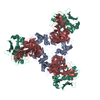
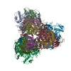
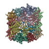
| Deposited unit | 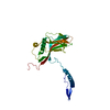
| ||||||||||
|---|---|---|---|---|---|---|---|---|---|---|---|
| 1 | x 12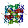
| ||||||||||
| Unit cell |
|
-Components
| #1: Protein | / Matrix protein Mass: 29378.559 Da / Num. of mol.: 1 Source method: isolated from a genetically manipulated source Source: (gene. exp.) Cydia pomonella granulosis virus (isolate Mexico/1963)Strain: isolate Mexico/1963 Production host: Cydia pomonella granulosis virus (isolate Mexican)References: UniProt: P87577 |
|---|---|
| #2: Water | ChemComp-HOH / Water Mass: 18.015 Da / Num. of mol.: 90 / Source method: isolated from a natural source / Formula: H2O Mass: 18.015 Da / Num. of mol.: 90 / Source method: isolated from a natural source / Formula: H2O |
-Experimental details
-Experiment
| Experiment | Method: ELECTRON CRYSTALLOGRAPHY |
|---|---|
| EM experiment | Aggregation state: 3D ARRAY / 3D reconstruction method: electron crystallography |
- Sample preparation
| Component | Name: Cydia pomonella granulosis virus (isolate Mexican) / Type: VIRUS / Entity ID: #1 / Source: NATURAL |
|---|---|
| Source (natural) | Organism: Cydia pomonella granulosis virus (isolate Mexican) |
| Details of virus | Empty: NO / Enveloped: NO / Isolate: OTHER / Type: VIRION |
| Buffer solution | pH: 7 |
| Specimen | Embedding applied: NO / Shadowing applied: NO / Staining applied: NO / Vitrification applied: YES |
| Vitrification | Cryogen name: ETHANE |
-Data collection
| Experimental equipment |  Model: Tecnai F20 / Image courtesy: FEI Company |
|---|---|
| Microscopy | Model: FEI TECNAI F20 |
| Electron gun | Electron source: FIELD EMISSION GUN / Accelerating voltage: 200 kV / Illumination mode: OTHER |
| Electron lens | Mode: DIFFRACTION / C2 aperture diameter: 5 µm / Alignment procedure: OTHER |
| Specimen holder | Cryogen: NITROGEN Specimen holder model: GATAN 626 SINGLE TILT LIQUID NITROGEN CRYO TRANSFER HOLDER Temperature (max): 100 K / Temperature (min): 93 K |
| Image recording | Average exposure time: 0.01 sec. / Electron dose: 4.7 e/Å2 / Film or detector model: OTHER / Num. of diffraction images: 32000 / Num. of grids imaged: 1 |
| Image scans | Sampling size: 50 µm / Width: 1536 / Height: 512 |
| EM diffraction | Camera length: 2580 mm / Tilt angle list: 0 |
| EM diffraction shell | Resolution: 10→1.55 Å / Fourier space coverage: 1 % / Multiplicity: 1 / Num. of structure factors: 1 / Phase residual: 1 ° |
| EM diffraction stats | Fourier space coverage: 100 % / High resolution: 1.6 Å / Num. of intensities measured: 9650574 / Num. of structure factors: 23422 / Phase error: 13.64 ° / Phase residual: 1 ° / Phase error rejection criteria: 0 / Rmerge: 0.093 / Rsym: 0.093 |
| Reflection | Biso Wilson estimate: 16.07 Å2 |
- Processing
| Software |
| ||||||||||||||||||||||||||||||||||||||||||||||||||||||||||||||||||||||
|---|---|---|---|---|---|---|---|---|---|---|---|---|---|---|---|---|---|---|---|---|---|---|---|---|---|---|---|---|---|---|---|---|---|---|---|---|---|---|---|---|---|---|---|---|---|---|---|---|---|---|---|---|---|---|---|---|---|---|---|---|---|---|---|---|---|---|---|---|---|---|---|
| EM software |
| ||||||||||||||||||||||||||||||||||||||||||||||||||||||||||||||||||||||
| EM 3D crystal entity | ∠α: 90 ° / ∠β: 90 ° / ∠γ: 90 ° / A: 103.2 Å / B: 103.2 Å / C: 103.2 Å / Space group name: I23 / Space group num: 197 | ||||||||||||||||||||||||||||||||||||||||||||||||||||||||||||||||||||||
| CTF correction | Details: Data reduction and structure solution using X-ray crystallography codes (CrystFEL, Phaser, cctbx.xfel, Coot) Type: NONE | ||||||||||||||||||||||||||||||||||||||||||||||||||||||||||||||||||||||
| 3D reconstruction | Resolution: 1.6 Å / Resolution method: DIFFRACTION PATTERN/LAYERLINES / Symmetry type: 3D CRYSTAL | ||||||||||||||||||||||||||||||||||||||||||||||||||||||||||||||||||||||
| Atomic model building | PDB-ID: 5G0Z Pdb chain-ID: A / Pdb chain residue range: 6-248 | ||||||||||||||||||||||||||||||||||||||||||||||||||||||||||||||||||||||
| Refinement | Resolution: 1.55→72.97 Å / SU ML: 0.1976 / Cross valid method: FREE R-VALUE / σ(F): 1.33 / Phase error: 21.1215 Stereochemistry target values: GeoStd + Monomer Library + CDL v1.2
| ||||||||||||||||||||||||||||||||||||||||||||||||||||||||||||||||||||||
| Solvent computation | Shrinkage radii: 0.9 Å / VDW probe radii: 1.11 Å / Solvent model: FLAT BULK SOLVENT MODEL | ||||||||||||||||||||||||||||||||||||||||||||||||||||||||||||||||||||||
| Displacement parameters | Biso mean: 19.29 Å2 | ||||||||||||||||||||||||||||||||||||||||||||||||||||||||||||||||||||||
| Refine LS restraints |
| ||||||||||||||||||||||||||||||||||||||||||||||||||||||||||||||||||||||
| LS refinement shell |
|