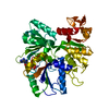
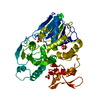
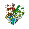
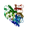
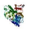
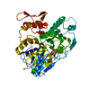
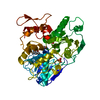
[English] 日本語
Yorodumi
- PDB-6s0j: Structure of Zika virus NS3 helicase in complex with ADP-MgF3(H2O)- -

+
Open data


ID or keywords:

Loading...

-
Basic information

Entry
Database: PDB / ID: 6s0j
TitleStructure of Zika virus NS3 helicase in complex with ADP-MgF3(H2O)-
ComponentsGenome polyprotein
KeywordsHYDROLASE / NS3 helicase
Function / homology
Function and homology information


symbiont-mediated suppression of host JAK-STAT cascade via inhibition of host TYK2 activity / flavivirin / symbiont-mediated suppression of host JAK-STAT cascade via inhibition of STAT2 activity / symbiont-mediated suppression of host JAK-STAT cascade via inhibition of STAT1 activity / negative regulation of innate immune response / ribonucleoside triphosphate phosphatase activity / viral capsid / nucleoside-triphosphate phosphatase / double-stranded RNA binding / 4 iron, 4 sulfur cluster binding ...symbiont-mediated suppression of host JAK-STAT cascade via inhibition of host TYK2 activity / flavivirin / symbiont-mediated suppression of host JAK-STAT cascade via inhibition of STAT2 activity / symbiont-mediated suppression of host JAK-STAT cascade via inhibition of STAT1 activity / negative regulation of innate immune response / ribonucleoside triphosphate phosphatase activity / viral capsid / nucleoside-triphosphate phosphatase / double-stranded RNA binding / 4 iron, 4 sulfur cluster binding / mRNA (guanine-N7)-methyltransferase / methyltransferase cap1 / clathrin-dependent endocytosis of virus by host cell / mRNA (nucleoside-2'-O-)-methyltransferase activity / mRNA 5'-cap (guanine-N7-)-methyltransferase activity / RNA helicase activity / molecular adaptor activity / host cell endoplasmic reticulum membrane / host cell perinuclear region of cytoplasm / protein dimerization activity / RNA helicase / induction by virus of host autophagy / symbiont entry into host cell / RNA-directed RNA polymerase / viral RNA genome replication / RNA-dependent RNA polymerase activity / serine-type endopeptidase activity / centrosome / fusion of virus membrane with host endosome membrane / viral envelope / lipid binding / symbiont-mediated suppression of host type I interferon-mediated signaling pathway / host cell nucleus / virion attachment to host cell / GTP binding / virion membrane / structural molecule activity / ATP hydrolysis activity / proteolysis / extracellular region / ATP binding / membrane / metal ion binding
Similarity search - Function
: / Flavivirus envelope glycoprotein E, stem/anchor domain / RNA-directed RNA polymerase, thumb domain, Flavivirus / Flavivirus RNA-directed RNA polymerase, thumb domain / Flavivirus capsid protein C superfamily / : / Flavivirus NS3 helicase, C-terminal helical domain / Flavivirus non-structural protein NS2B / Genome polyprotein, Flavivirus / Flavivirus non-structural protein NS4A ...: / Flavivirus envelope glycoprotein E, stem/anchor domain / RNA-directed RNA polymerase, thumb domain, Flavivirus / Flavivirus RNA-directed RNA polymerase, thumb domain / Flavivirus capsid protein C superfamily / : / Flavivirus NS3 helicase, C-terminal helical domain / Flavivirus non-structural protein NS2B / Genome polyprotein, Flavivirus / Flavivirus non-structural protein NS4A / Flavivirus non-structural protein NS2B / Flavivirus capsid protein C / Flavivirus non-structural protein NS4B / mRNA cap 0/1 methyltransferase / Flavivirus capsid protein C / Flavivirus non-structural protein NS4B / Flavivirus non-structural protein NS4A / Flavivirus NS2B domain profile. / mRNA cap 0 and cap 1 methyltransferase (EC 2.1.1.56 and EC 2.1.1.57) domain profile. / Flavivirus non-structural protein NS2A / Flavivirus non-structural protein NS2A / Envelope glycoprotein M, flavivirus / Flavivirus envelope glycoprotein M / Flavivirus NS3, petidase S7 / Peptidase S7, Flavivirus NS3 serine protease / Flavivirus NS3 protease (NS3pro) domain profile. / RNA-directed RNA polymerase, flavivirus / Flavivirus RNA-directed RNA polymerase, fingers and palm domains / Flavivirus non-structural Protein NS1 / Flavivirus non-structural protein NS1 / Envelope glycoprotein M superfamily, flavivirus / Flavivirus polyprotein propeptide / Flavivirus polyprotein propeptide superfamily / Flavivirus polyprotein propeptide / Flaviviral glycoprotein E, central domain, subdomain 1 / Flaviviral glycoprotein E, central domain, subdomain 2 / Flavivirus envelope glycoprotein E, Stem/Anchor domain / Flavivirus glycoprotein E, immunoglobulin-like domain / Flavivirus envelope glycoprotein E, Stem/Anchor domain superfamily / Flavivirus glycoprotein, immunoglobulin-like domain / Flavivirus glycoprotein central and dimerisation domain / Flavivirus glycoprotein, central and dimerisation domains / Ribosomal RNA methyltransferase, FtsJ domain / FtsJ-like methyltransferase / Flavivirus/Alphavirus glycoprotein, immunoglobulin-like domain superfamily / Flavivirus glycoprotein, central and dimerisation domain superfamily / Flaviviral glycoprotein E, dimerisation domain / DEAD box, Flavivirus / Flavivirus DEAD domain / helicase superfamily c-terminal domain / Immunoglobulin E-set / Superfamilies 1 and 2 helicase C-terminal domain profile. / Superfamilies 1 and 2 helicase ATP-binding type-1 domain profile. / DEAD-like helicases superfamily / Helicase, C-terminal / Helicase superfamily 1/2, ATP-binding domain / RNA-directed RNA polymerase, catalytic domain / RdRp of positive ssRNA viruses catalytic domain profile. / S-adenosyl-L-methionine-dependent methyltransferase superfamily / Peptidase S1, PA clan / DNA/RNA polymerase superfamily / P-loop containing nucleoside triphosphate hydrolase
Similarity search - Domain/homology
ADENOSINE-5'-DIPHOSPHATE / trifluoromagnesate monohydrate / Genome polyprotein / Genome polyprotein
Similarity search - Component
Biological speciesZika virus
MethodX-RAY DIFFRACTION / SYNCHROTRON / MOLECULAR REPLACEMENT / Resolution: 1.5 Å
AuthorsGe, M. / Molt Jr., R.W. / Jenkins, H.T. / Blackburn, G.M. / Jin, Y. / Antson, A.A.
Funding support United Kingdom, 2items
OrganizationGrant numberCountry
Wellcome TrustWT098230 United Kingdom
Wellcome TrustWT101528 United Kingdom
CitationJournal: To Be Published
Title: New MFx transition state analog reveals the molecular mechanism of ATP hydrolysis by the Zika virus NS3 helicase
Authors: Ge, M. / Molt Jr., R.W. / Jenkins, H.T. / Blackburn, G.M. / Jin, Y. / Antson, A.A.
History
DepositionJun 16, 2019Deposition site: PDBE / Processing site: PDBE
Revision 1.0Jul 15, 2020Provider: repository / Type: Initial release
Revision 1.1Jan 24, 2024Group: Data collection / Database references ...Data collection / Database references / Derived calculations / Refinement description
Category: chem_comp_atom / chem_comp_bond ...chem_comp_atom / chem_comp_bond / database_2 / pdbx_initial_refinement_model / pdbx_struct_conn_angle / struct_conn
Item: _database_2.pdbx_DOI / _database_2.pdbx_database_accession ..._database_2.pdbx_DOI / _database_2.pdbx_database_accession / _pdbx_struct_conn_angle.ptnr1_auth_comp_id / _pdbx_struct_conn_angle.ptnr1_auth_seq_id / _pdbx_struct_conn_angle.ptnr1_label_asym_id / _pdbx_struct_conn_angle.ptnr1_label_atom_id / _pdbx_struct_conn_angle.ptnr1_label_comp_id / _pdbx_struct_conn_angle.ptnr3_auth_comp_id / _pdbx_struct_conn_angle.ptnr3_auth_seq_id / _pdbx_struct_conn_angle.ptnr3_label_asym_id / _pdbx_struct_conn_angle.ptnr3_label_atom_id / _pdbx_struct_conn_angle.ptnr3_label_comp_id / _pdbx_struct_conn_angle.value / _struct_conn.pdbx_dist_value / _struct_conn.ptnr2_auth_comp_id / _struct_conn.ptnr2_auth_seq_id / _struct_conn.ptnr2_label_asym_id / _struct_conn.ptnr2_label_atom_id / _struct_conn.ptnr2_label_comp_id

-
Structure visualization

Structure viewerMolecule:
MolmilJmol/JSmol

Downloads & links

-
Assembly

Deposited unit
A: Genome polyprotein
hetero molecules


Theoretical massNumber of molelcules
Total (without water)51,2924
Polymers50,7431
Non-polymers5493
Water8,341463
1


  • Idetical with deposited unit
  • defined by author&software
  • Evidence: gel filtration
TypeNameSymmetry operationNumber
identity operation1_555x,y,z1
Buried area730 Å2
ΔGint-18 kcal/mol
Surface area19420 Å2
MethodPISA
Unit cell
Length a, b, c (Å)52.943, 69.604, 57.751
Angle α, β, γ (deg.)90.000, 94.400, 90.000
Int Tables number4
Space group name H-MP1211

-
Components

#1: Protein Genome polyprotein


Mass: 50742.871 Da / Num. of mol.: 1
Source method: isolated from a genetically manipulated source
Source: (gene. exp.) Zika virus (strain Mr 766) / Strain: ZIKV/H.sapiens/Brazil/PE243/2015 / Production host: Escherichia coli (E. coli) / References: UniProt: A0A160JCU6, UniProt: A0A024B7W1*PLUS
#2: Chemical ChemComp-KQB / trifluoromagnesate monohydrate


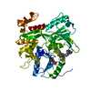
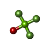
Mass: 97.300 Da / Num. of mol.: 1 / Source method: obtained synthetically / Formula: F3MgO / Feature type: SUBJECT OF INVESTIGATION
#3: Chemical ChemComp-MG / MAGNESIUM ION


Mass: 24.305 Da / Num. of mol.: 1 / Source method: obtained synthetically / Formula: Mg / Feature type: SUBJECT OF INVESTIGATION
#4: Chemical ChemComp-ADP / ADENOSINE-5'-DIPHOSPHATE / Adenosine diphosphate


Mass: 427.201 Da / Num. of mol.: 1 / Source method: obtained synthetically / Formula: C10H15N5O10P2 / Feature type: SUBJECT OF INVESTIGATION / Comment: ADP, energy-carrying molecule*YM
#5: Water ChemComp-HOH / water / Water


Mass: 18.015 Da / Num. of mol.: 463 / Source method: isolated from a natural source / Formula: H2O
Has ligand of interestY

-
Experimental details

-
Experiment

ExperimentMethod: X-RAY DIFFRACTION / Number of used crystals: 1

-
Sample preparation

CrystalDensity Matthews: 2.09 Å3/Da / Density % sol: 41.17 %
Crystal growTemperature: 293 K / Method: vapor diffusion
Details: 15% PEG3350, 0.16 M sodium citrate and 9% ethylene glycol

-
Data collection

DiffractionMean temperature: 100 K / Serial crystal experiment: N
Diffraction sourceSource: SYNCHROTRON / Site: Diamond / Beamline: I03 / Wavelength: 0.9795 Å
DetectorType: DECTRIS PILATUS3 6M / Detector: PIXEL / Date: May 23, 2017
RadiationProtocol: SINGLE WAVELENGTH / Monochromatic (M) / Laue (L): M / Scattering type: x-ray
Radiation wavelengthWavelength: 0.9795 Å / Relative weight: 1
ReflectionResolution: 1.5→69.6 Å / Num. obs: 66685 / % possible obs: 99.7 % / Redundancy: 2.9 % / CC1/2: 0.998 / Rmerge(I) obs: 0.044 / Rpim(I) all: 0.031 / Rrim(I) all: 0.054 / Net I/σ(I): 10.1 / Num. measured all: 194771 / Scaling rejects: 17
Reflection shellResolution: 1.5→1.53 Å / Redundancy: 2.9 % / Rmerge(I) obs: 0.988 / Mean I/σ(I) obs: 1.2 / Num. unique obs: 3292 / CC1/2: 0.419 / Rpim(I) all: 0.691 / Rrim(I) all: 1.211 / % possible all: 99

-
Processing

Software
NameVersionClassification
REFMAC5.8.0238refinement
Aimlessdata scaling
DIALSdata reduction
PHASERphasing
PDB_EXTRACTdata extraction
RefinementMethod to determine structure: MOLECULAR REPLACEMENT
Starting model: 2JLQ
Resolution: 1.5→57.65 Å / Cor.coef. Fo:Fc: 0.977 / Cor.coef. Fo:Fc free: 0.962 / Cross valid method: THROUGHOUT
RfactorNum. reflection% reflectionSelection details
Rfree0.1888 3285 4.9 %RANDOM
Rwork0.145 ---
obs0.1472 63340 99.54 %-
Displacement parametersBiso max: 81.7 Å2 / Biso mean: 26.885 Å2 / Biso min: 14.35 Å2
Refinement stepCycle: final / Resolution: 1.5→57.65 Å
ProteinNucleic acidLigandSolventTotal
Num. atoms3372 0 33 463 3868
Biso mean--31.39 44.51 -
Num. residues----433
Refine LS restraints
Refine-IDTypeDev idealDev ideal targetNumber
X-RAY DIFFRACTIONr_bond_refined_d0.010.0133563
X-RAY DIFFRACTIONr_bond_other_d0.0020.0173250
X-RAY DIFFRACTIONr_angle_refined_deg1.5861.6534858
X-RAY DIFFRACTIONr_angle_other_deg1.4511.5787500
X-RAY DIFFRACTIONr_dihedral_angle_1_deg6.735453
X-RAY DIFFRACTIONr_dihedral_angle_2_deg28.46620.266188
X-RAY DIFFRACTIONr_dihedral_angle_3_deg12.39115572
X-RAY DIFFRACTIONr_dihedral_angle_4_deg16.3231533
X-RAY DIFFRACTIONr_chiral_restr0.0780.2478
X-RAY DIFFRACTIONr_gen_planes_refined0.0080.024076
X-RAY DIFFRACTIONr_gen_planes_other0.0010.02795
X-RAY DIFFRACTIONr_rigid_bond_restr2.24836813
LS refinement shellHighest resolution: 1.5 Å

+
About Yorodumi

-
News

-
Feb 9, 2022. New format data for meta-information of EMDB entries

New format data for meta-information of EMDB entries

  • Version 3 of the EMDB header file is now the official format.
  • The previous official version 1.9 will be removed from the archive.

Related info.:EMDB header

External links:wwPDB to switch to version 3 of the EMDB data model

-
Aug 12, 2020. Covid-19 info

Covid-19 info

URL: https://pdbj.org/emnavi/covid19.php

New page: Covid-19 featured information page in EM Navigator.

Related info.:Covid-19 info / Mar 5, 2020. Novel coronavirus structure data

+
Mar 5, 2020. Novel coronavirus structure data

Novel coronavirus structure data

Related info.:Yorodumi Speices / Aug 12, 2020. Covid-19 info

External links:COVID-19 featured content - PDBj / Molecule of the Month (242):Coronavirus Proteases

+
Jan 31, 2019. EMDB accession codes are about to change! (news from PDBe EMDB page)

EMDB accession codes are about to change! (news from PDBe EMDB page)

  • The allocation of 4 digits for EMDB accession codes will soon come to an end. Whilst these codes will remain in use, new EMDB accession codes will include an additional digit and will expand incrementally as the available range of codes is exhausted. The current 4-digit format prefixed with “EMD-” (i.e. EMD-XXXX) will advance to a 5-digit format (i.e. EMD-XXXXX), and so on. It is currently estimated that the 4-digit codes will be depleted around Spring 2019, at which point the 5-digit format will come into force.
  • The EM Navigator/Yorodumi systems omit the EMD- prefix.

Related info.:Q: What is EMD? / ID/Accession-code notation in Yorodumi/EM Navigator

External links:EMDB Accession Codes are Changing Soon! / Contact to PDBj

+
Jul 12, 2017. Major update of PDB

Major update of PDB

  • wwPDB released updated PDB data conforming to the new PDBx/mmCIF dictionary.
  • This is a major update changing the version number from 4 to 5, and with Remediation, in which all the entries are updated.
  • In this update, many items about electron microscopy experimental information are reorganized (e.g. em_software).
  • Now, EM Navigator and Yorodumi are based on the updated data.

External links:wwPDB Remediation / Enriched Model Files Conforming to OneDep Data Standards Now Available in the PDB FTP Archive

-
Yorodumi

Thousand views of thousand structures

  • Yorodumi is a browser for structure data from EMDB, PDB, SASBDB, etc.
  • This page is also the successor to EM Navigator detail page, and also detail information page/front-end page for Omokage search.
  • The word "yorodu" (or yorozu) is an old Japanese word meaning "ten thousand". "mi" (miru) is to see.

Related info.:EMDB / PDB / SASBDB / Comparison of 3 databanks / Yorodumi Search / Aug 31, 2016. New EM Navigator & Yorodumi / Yorodumi Papers / Jmol/JSmol / Function and homology information / Changes in new EM Navigator and Yorodumi

Read more