 Movie
Movie Controller

[English] 日本語
 Yorodumi
- PDB-6rnk: Crystal structure of a humanized (K18E, K269N) rat succinate rece... -
+ Open data

- Basic information
| Entry | Database: PDB / ID: 6rnk | ||||||
|---|---|---|---|---|---|---|---|
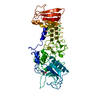
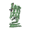
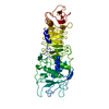
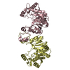
| Title | Crystal structure of a humanized (K18E, K269N) rat succinate receptor SUCNR1 (GPR91) in complex with a nanobody and antagonist NF-56-EJ40. | ||||||
 Components Components |
| ||||||
 Keywords Keywords |  MEMBRANE PROTEIN / SUCNR1 / GPR91 / GPCR / G-Protein coupled receptor / Nanobody / Antagonist / Succinate / Complex MEMBRANE PROTEIN / SUCNR1 / GPR91 / GPCR / G-Protein coupled receptor / Nanobody / Antagonist / Succinate / Complex | ||||||
| Function / homology |  Function and homology information Function and homology informationClass A/1 (Rhodopsin-like receptors) / regulation of angiotensin metabolic process / renin secretion into blood stream / positive regulation of chemotaxis / macrophage activation involved in immune response / G alpha (i) signalling events / G protein-coupled receptor activity / positive regulation of inflammatory response / response to calcium ion / glucose homeostasis ...Class A/1 (Rhodopsin-like receptors) / regulation of angiotensin metabolic process / renin secretion into blood stream / positive regulation of chemotaxis / macrophage activation involved in immune response / G alpha (i) signalling events / G protein-coupled receptor activity / positive regulation of inflammatory response / response to calcium ion / glucose homeostasis / signaling receptor activity / G protein-coupled receptor signaling pathway / plasma membraneSimilarity search - Function | ||||||
| Biological species |  Rattus norvegicus (Norway rat)Vicugna pacos (alpaca) Rattus norvegicus (Norway rat)Vicugna pacos (alpaca) | ||||||
| Method | X-RAY DIFFRACTION / SYNCHROTRON / MOLECULAR REPLACEMENT / Resolution: 1.94 Å | ||||||
 Authors Authors | Haffke, M. / Jaakola, V.-P. | ||||||
 Citation Citation | Journal: Nature / Year: 2019 Title: Structural basis of species-selective antagonist binding to the succinate receptor. Authors: Haffke, M. / Fehlmann, D. / Rummel, G. / Boivineau, J. / Duckely, M. / Gommermann, N. / Cotesta, S. / Sirockin, F. / Freuler, F. / Littlewood-Evans, A. / Kaupmann, K. / Jaakola, V.P. | ||||||
| History |
|
- Structure visualization
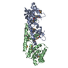
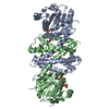
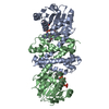
| Structure viewer | Molecule: MolmilJmol/JSmol |
|---|
- Downloads & links
-Download
| PDBx/mmCIF format | 6rnk.cif.gz | 207.3 KB | Display | PDBx/mmCIF format |
|---|---|---|---|---|
| PDB format | pdb6rnk.ent.gz | 163.1 KB | Display | PDB format |
| PDBx/mmJSON format | 6rnk.json.gz | Tree view | PDBx/mmJSON format | |
| Others |  Other downloads Other downloads |
-Validation report
| Arichive directory | https://data.pdbj.org/pub/pdb/validation_reports/rn/6rnkftp://data.pdbj.org/pub/pdb/validation_reports/rn/6rnk | HTTPS FTP |
|---|
-Related structure data
| Related structure data | 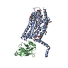 6ibbSC S: Starting model for refinement C: citing same article ( |
|---|---|
| Similar structure data |


-Links
 PDBj
PDBj


- Assembly
| Deposited unit | 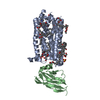
| |||||||||
|---|---|---|---|---|---|---|---|---|---|---|
| 1 |
| |||||||||
| Unit cell |
| |||||||||
| Components on special symmetry positions |
|
-Components
-Protein / Antibody , 2 types, 2 molecules AB
| #1: Protein | Mass: 39827.676 Da / Num. of mol.: 1 / Mutation: K18E, K269N Source method: isolated from a genetically manipulated source Source: (gene. exp.) Rattus norvegicus (Norway rat) / Gene: Sucnr1, Gpr91 / Production host:   Spodoptera frugiperda (fall armyworm) / References: UniProt: Q6IYF9 Spodoptera frugiperda (fall armyworm) / References: UniProt: Q6IYF9 |
|---|---|
| #2: Antibody | Mass: 15527.099 Da / Num. of mol.: 1 Source method: isolated from a genetically manipulated source Source: (gene. exp.) Vicugna pacos (alpaca) / Production host: Spodoptera frugiperda (fall armyworm) |
-Non-polymers , 5 types, 258 molecules 


| #3: Chemical | ChemComp-KAZ /  Mass: 443.537 Da / Num. of mol.: 1 / Source method: obtained synthetically / Formula: C27H29N3O3 Mass: 443.537 Da / Num. of mol.: 1 / Source method: obtained synthetically / Formula: C27H29N3O3 | ||||||
|---|---|---|---|---|---|---|---|
| #4: Chemical | ChemComp-OLC / (  Mass: 356.540 Da / Num. of mol.: 13 / Source method: obtained synthetically / Formula: C21H40O4 Mass: 356.540 Da / Num. of mol.: 13 / Source method: obtained synthetically / Formula: C21H40O4#5: Chemical | ChemComp-GOL / | Glycerol Mass: 92.094 Da / Num. of mol.: 1 / Source method: obtained synthetically / Formula: C3H8O3 Mass: 92.094 Da / Num. of mol.: 1 / Source method: obtained synthetically / Formula: C3H8O3#6: Chemical | ChemComp-SO4 / | Sulfate Mass: 96.063 Da / Num. of mol.: 1 / Source method: obtained synthetically / Formula: SO4 Mass: 96.063 Da / Num. of mol.: 1 / Source method: obtained synthetically / Formula: SO4#7: Water | ChemComp-HOH / | WaterMass: 18.015 Da / Num. of mol.: 242 / Source method: isolated from a natural source / Formula: H2O |
-Experimental details
-Experiment
| Experiment | Method: X-RAY DIFFRACTION / Number of used crystals: 1 |
|---|
- Sample preparation
| Crystal | Density Matthews: 3.36 Å3/Da / Density % sol: 63.5 % |
|---|---|
| Crystal grow | Temperature: 293.15 K / Method: lipidic cubic phase / pH: 7 Details: 50 mM ADA, pH 7.0, 28% PEG MME 550, 0.55 M (NH4)2SO4, 100-400 microM compound 3, 1-4 % (v/v) DMSO |
-Data collection
| Diffraction | Mean temperature: 100 K / Serial crystal experiment: N |
|---|---|
| Diffraction source | Source: SYNCHROTRON / Site: SLS  / Beamline: X06SA / Wavelength: 1.00002 Å / Beamline: X06SA / Wavelength: 1.00002 Å |
| Detector | Type: DECTRIS EIGER X 16M / Detector: PIXEL / Date: Apr 29, 2019 |
| Radiation | Protocol: SINGLE WAVELENGTH / Monochromatic (M) / Laue (L): M / Scattering type: x-ray |
| Radiation wavelength | Wavelength: 1.00002 Å / Relative weight: 1 |
| Reflection | Resolution: 1.43→75.47 Å / Num. obs: 88565 / % possible obs: 67 % / Redundancy: 6.5 % / Biso Wilson estimate: 34.46 Å2 / CC1/2: 0.998 / Rmerge(I) obs: 0.213 / Rpim(I) all: 0.091 / Rrim(I) all: 0.232 / Net I/σ(I): 4.8 / Num. measured all: 575822 / Scaling rejects: 8 |
| Reflection shell | Resolution: 1.43→1.51 Å / Redundancy: 3.4 % / Rmerge(I) obs: 8.756 / Num. measured all: 5597 / Num. unique obs: 1652 / Rpim(I) all: 5.465 / Rrim(I) all: 10.378 / Net I/σ(I) obs: 0.2 / % possible all: 8.6 |
- Processing
| Software |
| ||||||||||||||||||||||||||||||||||||||||||||||||||||||||||||||||||||||||||||||||||||||||||||||||||||||||||||
|---|---|---|---|---|---|---|---|---|---|---|---|---|---|---|---|---|---|---|---|---|---|---|---|---|---|---|---|---|---|---|---|---|---|---|---|---|---|---|---|---|---|---|---|---|---|---|---|---|---|---|---|---|---|---|---|---|---|---|---|---|---|---|---|---|---|---|---|---|---|---|---|---|---|---|---|---|---|---|---|---|---|---|---|---|---|---|---|---|---|---|---|---|---|---|---|---|---|---|---|---|---|---|---|---|---|---|---|---|---|
| Refinement | Method to determine structure: MOLECULAR REPLACEMENT Starting model: 6IBB Resolution: 1.94→75.47 Å / Cor.coef. Fo:Fc: 0.953 / Cor.coef. Fo:Fc free: 0.932 / SU R Cruickshank DPI: 0.145 / Cross valid method: THROUGHOUT / σ(F): 0 / SU R Blow DPI: 0.144 / SU Rfree Blow DPI: 0.128 / SU Rfree Cruickshank DPI: 0.129
| ||||||||||||||||||||||||||||||||||||||||||||||||||||||||||||||||||||||||||||||||||||||||||||||||||||||||||||
| Displacement parameters | Biso max: 154.62 Å2 / Biso mean: 45.42 Å2 / Biso min: 21.61 Å2
| ||||||||||||||||||||||||||||||||||||||||||||||||||||||||||||||||||||||||||||||||||||||||||||||||||||||||||||
| Refine analyze | Luzzati coordinate error obs: 0.23 Å | ||||||||||||||||||||||||||||||||||||||||||||||||||||||||||||||||||||||||||||||||||||||||||||||||||||||||||||
| Refinement step | Cycle: final / Resolution: 1.94→75.47 Å
| ||||||||||||||||||||||||||||||||||||||||||||||||||||||||||||||||||||||||||||||||||||||||||||||||||||||||||||
| Refine LS restraints |
| ||||||||||||||||||||||||||||||||||||||||||||||||||||||||||||||||||||||||||||||||||||||||||||||||||||||||||||
| LS refinement shell | Resolution: 1.94→2.03 Å / Rfactor Rfree error: 0 / Total num. of bins used: 50
| ||||||||||||||||||||||||||||||||||||||||||||||||||||||||||||||||||||||||||||||||||||||||||||||||||||||||||||
| Refinement TLS params. | Method: refined / Refine-ID: X-RAY DIFFRACTION
| ||||||||||||||||||||||||||||||||||||||||||||||||||||||||||||||||||||||||||||||||||||||||||||||||||||||||||||
| Refinement TLS group |
|
