 Movie
Movie Controller


+ Open data

- Basic information
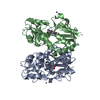
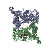
| Entry | Database: PDB / ID: 6ras | ||||||
|---|---|---|---|---|---|---|---|
| Title | Pmar-Lig_Pre. | ||||||
 Components Components |
| ||||||
 Keywords Keywords |  DNA BINDING PROTEIN / DNA ligase / ATP-dependent / ligase-DNA co-crystal structure / determinants in DNA binding DNA BINDING PROTEIN / DNA ligase / ATP-dependent / ligase-DNA co-crystal structure / determinants in DNA binding | ||||||
| Function / homology |  Function and homology informationDNA ligase (ATP) activity / DNA recombination / DNA replication / DNA repair / ATP binding / metal ion binding Function and homology informationDNA ligase (ATP) activity / DNA recombination / DNA replication / DNA repair / ATP binding / metal ion bindingSimilarity search - Function | ||||||
| Biological species |  Prochlorococcus marinus str. MIT 9302 (bacteria) Prochlorococcus marinus str. MIT 9302 (bacteria)DNA launch vector pDE-GFP2 (others) | ||||||
| Method | X-RAY DIFFRACTION / SYNCHROTRON / MOLECULAR REPLACEMENT / Resolution: 2.75 Å | ||||||
 Authors Authors | Leiros, H.K.S. / Williamson, A. | ||||||
| Funding support |  Norway, 1items Norway, 1items
| ||||||
 Citation Citation | Journal: Nucleic Acids Res. / Year: 2019 Title: Structural intermediates of a DNA-ligase complex illuminate the role of the catalytic metal ion and mechanism of phosphodiester bond formation. Authors: Williamson, A. / Leiros, H.S. | ||||||
| History |
|
- Structure visualization
| Structure viewer | Molecule: MolmilJmol/JSmol |
|---|
- Downloads & links
-Download
| PDBx/mmCIF format | 6ras.cif.gz | 221.2 KB | Display | PDBx/mmCIF format |
|---|---|---|---|---|
| PDB format | pdb6ras.ent.gz | 173.8 KB | Display | PDB format |
| PDBx/mmJSON format | 6ras.json.gz | Tree view | PDBx/mmJSON format | |
| Others |  Other downloads Other downloads |
-Validation report
| Arichive directory | https://data.pdbj.org/pub/pdb/validation_reports/ra/6rasftp://data.pdbj.org/pub/pdb/validation_reports/ra/6ras | HTTPS FTP |
|---|
-Related structure data


-Links
 PDBj
PDBj


- Assembly
| Deposited unit | 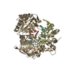
| ||||||||
|---|---|---|---|---|---|---|---|---|---|
| 1 |
| ||||||||
| Unit cell |
|
-Components
-DNA chain , 3 types, 3 molecules BCD
| #1: DNA chain | Mass: 6479.183 Da / Num. of mol.: 1 / Source method: obtained synthetically / Source: (synth.) DNA launch vector pDE-GFP2 (others) |
|---|---|
| #2: DNA chain | Mass: 3035.007 Da / Num. of mol.: 1 / Source method: obtained synthetically Details: There is a break in the chain between nt31 and nt32. Source: (synth.) DNA launch vector pDE-GFP2 (others) |
| #3: DNA chain | Mass: 3342.212 Da / Num. of mol.: 1 / Source method: obtained synthetically / Source: (synth.) DNA launch vector pDE-GFP2 (others) |
-Protein , 1 types, 1 molecules I
| #4: Protein | Mass: 50886.648 Da / Num. of mol.: 1 Source method: isolated from a genetically manipulated source Details: This is a R120 A mutant Source: (gene. exp.) Prochlorococcus marinus str. MIT 9302 (bacteria)Gene: EU96_0746 / Production host: Prochlorococcus marinus (bacteria) / References: UniProt: A0A0A2ACP7 |
|---|
-Non-polymers , 4 types, 201 molecules 


| #5: Chemical | Polyethylene glycol Mass: 150.173 Da / Num. of mol.: 2 / Source method: obtained synthetically / Formula: C6H14O4 Mass: 150.173 Da / Num. of mol.: 2 / Source method: obtained synthetically / Formula: C6H14O4#6: Chemical | ChemComp-AMP / | Adenosine monophosphate Mass: 347.221 Da / Num. of mol.: 1 / Source method: obtained synthetically / Formula: C10H14N5O7P / Feature type: SUBJECT OF INVESTIGATION / Comment: AMP*YM Mass: 347.221 Da / Num. of mol.: 1 / Source method: obtained synthetically / Formula: C10H14N5O7P / Feature type: SUBJECT OF INVESTIGATION / Comment: AMP*YM#7: Chemical | Sulfate Mass: 96.063 Da / Num. of mol.: 2 / Source method: obtained synthetically / Formula: SO4 Mass: 96.063 Da / Num. of mol.: 2 / Source method: obtained synthetically / Formula: SO4#8: Water | ChemComp-HOH / | WaterMass: 18.015 Da / Num. of mol.: 196 / Source method: isolated from a natural source / Formula: H2O |
|---|
-Experimental details
-Experiment
| Experiment | Method: X-RAY DIFFRACTION / Number of used crystals: 1 |
|---|
- Sample preparation
| Crystal | Density Matthews: 3.54 Å3/Da / Density % sol: 65.28 % |
|---|---|
| Crystal grow | Temperature: 277 K / Method: vapor diffusion / Details: PEG, buffer |
-Data collection
| Diffraction | Mean temperature: 100 K / Serial crystal experiment: N |
|---|---|
| Diffraction source | Source: SYNCHROTRON / Site: BESSY  / Beamline: 14.2 / Wavelength: 0.9184 Å / Beamline: 14.2 / Wavelength: 0.9184 Å |
| Detector | Type: DECTRIS PILATUS3 S 6M / Detector: PIXEL / Date: Jul 4, 2018 |
| Radiation | Protocol: SINGLE WAVELENGTH / Monochromatic (M) / Laue (L): M / Scattering type: x-ray |
| Radiation wavelength | Wavelength: 0.9184 Å / Relative weight: 1 |
| Reflection | Resolution: 2.69→25 Å / Num. obs: 24531 / % possible obs: 99.4 % / Redundancy: 5.6 % / Biso Wilson estimate: 42.2 Å2 / CC1/2: 0.995 / Rmerge(I) obs: 0.129 / Rpim(I) all: 0.089 / Net I/av σ(I): 9.5 / Net I/σ(I): 9.5 |
| Reflection shell | Resolution: 2.69→2.82 Å / Rmerge(I) obs: 1.16 / Mean I/σ(I) obs: 1.3 / Num. unique obs: 3459 / CC1/2: 0.475 / Rpim(I) all: 0.602 / % possible all: 99.6 |
- Processing
| Software |
| ||||||||||||||||||||||||||||||||||||||||||||||||||||||||||||||||||||||
|---|---|---|---|---|---|---|---|---|---|---|---|---|---|---|---|---|---|---|---|---|---|---|---|---|---|---|---|---|---|---|---|---|---|---|---|---|---|---|---|---|---|---|---|---|---|---|---|---|---|---|---|---|---|---|---|---|---|---|---|---|---|---|---|---|---|---|---|---|---|---|---|
| Refinement | Method to determine structure: MOLECULAR REPLACEMENT Starting model: Home-made Resolution: 2.75→24.366 Å / SU ML: 0.38 / Cross valid method: THROUGHOUT / σ(F): 1.36 / Phase error: 25.79 / Stereochemistry target values: ML
| ||||||||||||||||||||||||||||||||||||||||||||||||||||||||||||||||||||||
| Solvent computation | Shrinkage radii: 0.9 Å / VDW probe radii: 1.11 Å / Solvent model: FLAT BULK SOLVENT MODEL | ||||||||||||||||||||||||||||||||||||||||||||||||||||||||||||||||||||||
| Refinement step | Cycle: LAST / Resolution: 2.75→24.366 Å
| ||||||||||||||||||||||||||||||||||||||||||||||||||||||||||||||||||||||
| Refine LS restraints |
| ||||||||||||||||||||||||||||||||||||||||||||||||||||||||||||||||||||||
| LS refinement shell |
|
