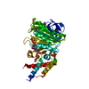
- PDB-6rae: Structural analysis of the Salmonella type III secretion system A... -
+
Open data
ID or keywords:
Loading...
-
Basic information
Entry
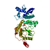
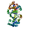
Database: PDB / ID: 6rae
Title
Structural analysis of the Salmonella type III secretion system ATPase InvC
Components
Secretory apparatus ATP synthase (Associated with virulence)
Keywords
HYDROLASE / Bacterial pathogenesis / Salmonella enterica / Type III secretion system (T3SS) / ATPase / Crystallography.
Function / homology
Function and homology information
protein-exporting ATPase activity / protein-secreting ATPase / type III protein secretion system complex / protein secretion by the type III secretion system / proton motive force-driven ATP synthesis / proton-transporting ATP synthase complex, catalytic core F(1) / proton transmembrane transport / proton-transporting ATPase activity, rotational mechanism / proton-transporting ATP synthase activity, rotational mechanism / ATP hydrolysis activity ...protein-exporting ATPase activity / protein-secreting ATPase / type III protein secretion system complex / protein secretion by the type III secretion system / proton motive force-driven ATP synthesis / proton-transporting ATP synthase complex, catalytic core F(1) / proton transmembrane transport / proton-transporting ATPase activity, rotational mechanism / proton-transporting ATP synthase activity, rotational mechanism / ATP hydrolysis activity / ATP binding / cytoplasm Similarity search - Function
ATPase, type III secretion system, FliI/YscN / T3SS EscN ATPase, C-terminal / T3SS EscN ATPase C-terminal domain / ATPase, F1/V1/A1 complex, alpha/beta subunit, N-terminal domain / ATP synthase alpha/beta family, beta-barrel domain / ATPase, alpha/beta subunit, nucleotide-binding domain, active site / ATP synthase alpha and beta subunits signature. / ATPase, F1/V1/A1 complex, alpha/beta subunit, nucleotide-binding domain / ATP synthase alpha/beta family, nucleotide-binding domain / ATPases associated with a variety of cellular activities ...ATPase, type III secretion system, FliI/YscN / T3SS EscN ATPase, C-terminal / T3SS EscN ATPase C-terminal domain / ATPase, F1/V1/A1 complex, alpha/beta subunit, N-terminal domain / ATP synthase alpha/beta family, beta-barrel domain / ATPase, alpha/beta subunit, nucleotide-binding domain, active site / ATP synthase alpha and beta subunits signature. / ATPase, F1/V1/A1 complex, alpha/beta subunit, nucleotide-binding domain / ATP synthase alpha/beta family, nucleotide-binding domain / ATPases associated with a variety of cellular activities / AAA+ ATPase domain / P-loop containing nucleoside triphosphate hydrolase Similarity search - Domain/homology
In the structure databanks used in Yorodumi, some data are registered as the other names, "COVID-19 virus" and "2019-nCoV". Here are the details of the virus and the list of structure data.
Jan 31, 2019. EMDB accession codes are about to change! (news from PDBe EMDB page)
EMDB accession codes are about to change! (news from PDBe EMDB page)
The allocation of 4 digits for EMDB accession codes will soon come to an end. Whilst these codes will remain in use, new EMDB accession codes will include an additional digit and will expand incrementally as the available range of codes is exhausted. The current 4-digit format prefixed with “EMD-” (i.e. EMD-XXXX) will advance to a 5-digit format (i.e. EMD-XXXXX), and so on. It is currently estimated that the 4-digit codes will be depleted around Spring 2019, at which point the 5-digit format will come into force.
The EM Navigator/Yorodumi systems omit the EMD- prefix.
Related info.:Q: What is EMD? / ID/Accession-code notation in Yorodumi/EM Navigator
Yorodumi is a browser for structure data from EMDB, PDB, SASBDB, etc.
This page is also the successor to EM Navigator detail page, and also detail information page/front-end page for Omokage search.
The word "yorodu" (or yorozu) is an old Japanese word meaning "ten thousand". "mi" (miru) is to see.
Related info.:EMDB / PDB / SASBDB / Comparison of 3 databanks / Yorodumi Search / Aug 31, 2016. New EM Navigator & Yorodumi / Yorodumi Papers / Jmol/JSmol / Function and homology information / Changes in new EM Navigator and Yorodumi
 Movie
Movie Controller

 Yorodumi
 Open data
 Basic information
 Components
 Keywords
 HYDROLASE / Bacterial pathogenesis /
  Function and homology information
 Authors
 Germany, 1items
  Citation
 Structure visualization
 Downloads & links
 Other downloads
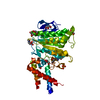


 PDBj
PDBj


 Assembly

 Mass: 92.094 Da / Num. of mol.: 1 / Source method: obtained synthetically / Formula: C3H8O3
 Mass: 35.453 Da / Num. of mol.: 2 / Source method: obtained synthetically / Formula: Cl
 Mass: 18.015 Da / Num. of mol.: 151 / Source method: isolated from a natural source / Formula: H2O
 Sample preparation
 Processing