 Movie
Movie Controller

[English] 日本語
 Yorodumi
- PDB-6r3d: Crystal structure of di-phosphorylated human CLK1 in complex with... -
+ Open data

- Basic information
| Entry | Database: PDB / ID: 6r3d | ||||||
|---|---|---|---|---|---|---|---|
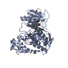
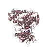
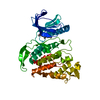
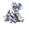
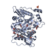
| Title | Crystal structure of di-phosphorylated human CLK1 in complex with 4-(6,7-dichloro-1H-indol-3-yl)pyrimidin-2-amine | ||||||
 Components Components | Dual specificity protein kinase CLK1 | ||||||
 Keywords Keywords |  SIGNALING PROTEIN / splicing / kinase / viral infection SIGNALING PROTEIN / splicing / kinase / viral infection | ||||||
| Function / homology |  Function and homology informationdual-specificity kinase / regulation of RNA splicing / protein serine/threonine/tyrosine kinase activity / non-membrane spanning protein tyrosine kinase activity / protein tyrosine kinase activity / phosphorylation / protein serine kinase activity / protein serine/threonine kinase activity / ATP binding / nucleus Function and homology informationdual-specificity kinase / regulation of RNA splicing / protein serine/threonine/tyrosine kinase activity / non-membrane spanning protein tyrosine kinase activity / protein tyrosine kinase activity / phosphorylation / protein serine kinase activity / protein serine/threonine kinase activity / ATP binding / nucleusSimilarity search - Function | ||||||
| Biological species |  Homo sapiens (human) Homo sapiens (human) | ||||||
| Method | X-RAY DIFFRACTION / SYNCHROTRON / MOLECULAR REPLACEMENT / Resolution: 1.85 Å | ||||||
 Authors Authors | Livnah, O. / Domovich, Y. | ||||||
 Citation Citation | Journal: To Be Published Title: Crystal structure of di-phosphorylated human CLK1 in complex with 4-(1-methyl-1H-indol-3-yl)pyrimidin-2-amine Authors: Livnah, O. / Domovich, Y. | ||||||
| History |
|
- Structure visualization
| Structure viewer | Molecule: MolmilJmol/JSmol |
|---|
- Downloads & links
-Download
| PDBx/mmCIF format | 6r3d.cif.gz | 84.6 KB | Display | PDBx/mmCIF format |
|---|---|---|---|---|
| PDB format | pdb6r3d.ent.gz | 62.4 KB | Display | PDB format |
| PDBx/mmJSON format | 6r3d.json.gz | Tree view | PDBx/mmJSON format | |
| Others |  Other downloads Other downloads |
-Validation report
| Arichive directory | https://data.pdbj.org/pub/pdb/validation_reports/r3/6r3dftp://data.pdbj.org/pub/pdb/validation_reports/r3/6r3d | HTTPS FTP |
|---|
-Related structure data
| Related structure data |  2vagS S: Starting model for refinement |
|---|---|
| Similar structure data |


-Links
 PDBj
PDBj- Assembly
| Deposited unit | 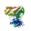
| ||||||||
|---|---|---|---|---|---|---|---|---|---|
| 1 |
| ||||||||
| Unit cell |
| ||||||||
| Components on special symmetry positions |
|
-Components
| #1: Protein | Mass: 39449.352 Da / Num. of mol.: 1 Source method: isolated from a genetically manipulated source Source: (gene. exp.) Homo sapiens (human) / Gene: CLK1, CLK / Production host:  Escherichia coli (E. coli) / References: UniProt: P49759, dual-specificity kinase Escherichia coli (E. coli) / References: UniProt: P49759, dual-specificity kinase |
|---|---|
| #2: Chemical | ChemComp-JQW /   Mass: 279.125 Da / Num. of mol.: 1 / Source method: obtained synthetically / Formula: C12H8Cl2N4 Mass: 279.125 Da / Num. of mol.: 1 / Source method: obtained synthetically / Formula: C12H8Cl2N4 |
| #3: Water | ChemComp-HOH / Water Mass: 18.015 Da / Num. of mol.: 97 / Source method: isolated from a natural source / Formula: H2O Mass: 18.015 Da / Num. of mol.: 97 / Source method: isolated from a natural source / Formula: H2O |
-Experimental details
-Experiment
| Experiment | Method: X-RAY DIFFRACTION / Number of used crystals: 1 |
|---|
- Sample preparation
| Crystal | Density Matthews: 2.46 Å3/Da / Density % sol: 49.92 % |
|---|---|
| Crystal grow | Temperature: 293 K / Method: vapor diffusion, sitting drop / pH: 6.5 / Details: 24% PEG 3350, 200mM MgCl2, 100mM BisTris (pH 6.5) |
-Data collection
| Diffraction | Mean temperature: 100 K / Serial crystal experiment: N |
|---|---|
| Diffraction source | Source: SYNCHROTRON / Site: ESRF  / Beamline: ID23-1 / Wavelength: 1 Å / Beamline: ID23-1 / Wavelength: 1 Å |
| Detector | Type: DECTRIS PILATUS3 S 6M / Detector: PIXEL / Date: Nov 22, 2016 |
| Radiation | Protocol: SINGLE WAVELENGTH / Monochromatic (M) / Laue (L): M / Scattering type: x-ray |
| Radiation wavelength | Wavelength: 1 Å / Relative weight: 1 |
| Reflection | Resolution: 1.85→47.52 Å / Num. obs: 35238 / % possible obs: 99.8 % / Redundancy: 6.2 % / Rsym value: 0.051 / Net I/σ(I): 15.8 |
| Reflection shell | Resolution: 1.85→1.898 Å / Num. unique obs: 2095 |
- Processing
| Software |
| ||||||||||||||||||||||||||||||||||||||||||||||||||||||||||||||||||||||||||||||||||||||||||||||||||||||||||||||||||||||||||||||||||||||||||||||||||||||||||||||||||||||||||||||||||||||
|---|---|---|---|---|---|---|---|---|---|---|---|---|---|---|---|---|---|---|---|---|---|---|---|---|---|---|---|---|---|---|---|---|---|---|---|---|---|---|---|---|---|---|---|---|---|---|---|---|---|---|---|---|---|---|---|---|---|---|---|---|---|---|---|---|---|---|---|---|---|---|---|---|---|---|---|---|---|---|---|---|---|---|---|---|---|---|---|---|---|---|---|---|---|---|---|---|---|---|---|---|---|---|---|---|---|---|---|---|---|---|---|---|---|---|---|---|---|---|---|---|---|---|---|---|---|---|---|---|---|---|---|---|---|---|---|---|---|---|---|---|---|---|---|---|---|---|---|---|---|---|---|---|---|---|---|---|---|---|---|---|---|---|---|---|---|---|---|---|---|---|---|---|---|---|---|---|---|---|---|---|---|---|---|
| Refinement | Method to determine structure: MOLECULAR REPLACEMENT Starting model: 2VAG Resolution: 1.85→47.52 Å / Cor.coef. Fo:Fc: 0.957 / Cor.coef. Fo:Fc free: 0.948 / SU B: 4.901 / SU ML: 0.137 / Cross valid method: THROUGHOUT / ESU R: 0.149 / ESU R Free: 0.145 / Details: HYDROGENS HAVE BEEN ADDED IN THE RIDING POSITIONS
| ||||||||||||||||||||||||||||||||||||||||||||||||||||||||||||||||||||||||||||||||||||||||||||||||||||||||||||||||||||||||||||||||||||||||||||||||||||||||||||||||||||||||||||||||||||||
| Solvent computation | Ion probe radii: 0.8 Å / Shrinkage radii: 0.8 Å / VDW probe radii: 1.2 Å | ||||||||||||||||||||||||||||||||||||||||||||||||||||||||||||||||||||||||||||||||||||||||||||||||||||||||||||||||||||||||||||||||||||||||||||||||||||||||||||||||||||||||||||||||||||||
| Displacement parameters | Biso mean: 45.963 Å2
| ||||||||||||||||||||||||||||||||||||||||||||||||||||||||||||||||||||||||||||||||||||||||||||||||||||||||||||||||||||||||||||||||||||||||||||||||||||||||||||||||||||||||||||||||||||||
| Refinement step | Cycle: LAST / Resolution: 1.85→47.52 Å
| ||||||||||||||||||||||||||||||||||||||||||||||||||||||||||||||||||||||||||||||||||||||||||||||||||||||||||||||||||||||||||||||||||||||||||||||||||||||||||||||||||||||||||||||||||||||
| Refine LS restraints |
|