 Movie
Movie Controller

[English] 日本語
 Yorodumi
- PDB-6r35: Structure of the LecB lectin from Pseudomonas aeruginosa strain P... -
+ Open data

- Basic information
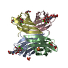
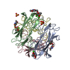
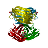
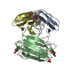
| Entry | Database: PDB / ID: 6r35 | |||||||||
|---|---|---|---|---|---|---|---|---|---|---|
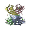
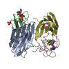
| Title | Structure of the LecB lectin from Pseudomonas aeruginosa strain PAO1 in complex with lewis x tetrasaccharide | |||||||||
 Components Components | Fucose-binding lectin PA-IIL | |||||||||
 Keywords Keywords | SUGAR BINDING PROTEIN /  Lectin / carbohydrate / Lewis x / LecB Lectin / carbohydrate / Lewis x / LecB | |||||||||
| Function / homology |  Function and homology information Function and homology informationsingle-species biofilm formation / carbohydrate binding / metal ion bindingSimilarity search - Function | |||||||||
| Biological species |  Pseudomonas aeruginosa PAO1 (bacteria) Pseudomonas aeruginosa PAO1 (bacteria) | |||||||||
| Method | X-RAY DIFFRACTION / SYNCHROTRON / MOLECULAR REPLACEMENT / Resolution: 1.8 Å | |||||||||
 Authors Authors | Lepsik, M. / Sommer, R. / Kuhaudomlarp, S. / Lelimousin, M. / Varrot, A. / Titz, A. / Imberty, A. | |||||||||
| Funding support |  France, 1items France, 1items
| |||||||||
 Citation Citation | Journal: Eur.J.Med.Chem. / Year: 2019 Title: Induction of rare conformation of oligosaccharide by binding to calcium-dependent bacterial lectin: X-ray crystallography and modelling study. Authors: Lepsik, M. / Sommer, R. / Kuhaudomlarp, S. / Lelimousin, M. / Paci, E. / Varrot, A. / Titz, A. / Imberty, A. | |||||||||
| History |
|
- Structure visualization
| Structure viewer | Molecule: MolmilJmol/JSmol |
|---|
- Downloads & links
-Download
| PDBx/mmCIF format | 6r35.cif.gz | 115.9 KB | Display | PDBx/mmCIF format |
|---|---|---|---|---|
| PDB format | pdb6r35.ent.gz | 88.1 KB | Display | PDB format |
| PDBx/mmJSON format | 6r35.json.gz | Tree view | PDBx/mmJSON format | |
| Others |  Other downloads Other downloads |
-Validation report
| Arichive directory | https://data.pdbj.org/pub/pdb/validation_reports/r3/6r35ftp://data.pdbj.org/pub/pdb/validation_reports/r3/6r35 | HTTPS FTP |
|---|
-Related structure data
| Related structure data |  5a70C  1w8hS C: citing same article ( S: Starting model for refinement |
|---|---|
| Similar structure data |
-Links
 PDBj
PDBj- Assembly
| Deposited unit | 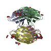
| ||||||||||||||||||||||||||||||||||||||||||||||||||||||||||||||||||||||||||||||||||||||||||||||||||
|---|---|---|---|---|---|---|---|---|---|---|---|---|---|---|---|---|---|---|---|---|---|---|---|---|---|---|---|---|---|---|---|---|---|---|---|---|---|---|---|---|---|---|---|---|---|---|---|---|---|---|---|---|---|---|---|---|---|---|---|---|---|---|---|---|---|---|---|---|---|---|---|---|---|---|---|---|---|---|---|---|---|---|---|---|---|---|---|---|---|---|---|---|---|---|---|---|---|---|---|
| 1 |
| ||||||||||||||||||||||||||||||||||||||||||||||||||||||||||||||||||||||||||||||||||||||||||||||||||
| Unit cell |
| ||||||||||||||||||||||||||||||||||||||||||||||||||||||||||||||||||||||||||||||||||||||||||||||||||
| Noncrystallographic symmetry (NCS) | NCS domain:
NCS domain segments: Component-ID: 0 / Beg auth comp-ID: ALA / Beg label comp-ID: ALA / End auth comp-ID: GLY / End label comp-ID: GLY / Refine code: 0 / Auth seq-ID: 1 - 114 / Label seq-ID: 1 - 114
NCS ensembles :
|
-Components
-Protein / Sugars , 2 types, 8 molecules DABC
| #1: Protein | Mass: 11734.707 Da / Num. of mol.: 4 Source method: isolated from a genetically manipulated source Details: The first methionine was cleaved. / Source: (gene. exp.) Pseudomonas aeruginosa PAO1 (bacteria) / Gene: lecB, PA3361 / Plasmid: pET25 / Production host: Escherichia coli BL21(DE3) (bacteria) / References: UniProt: Q9HYN5#2: Polysaccharide | alpha-L-fucopyranose-(1-3)-[beta-D-galactopyranose-(1-4)]2-acetamido-2-deoxy-beta-D-glucopyranose / Lewis X antigen / beta anomer 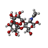  , Oligosaccharide / Class: Antigen / Mass: 529.490 Da / Num. of mol.: 4 , Oligosaccharide / Class: Antigen / Mass: 529.490 Da / Num. of mol.: 4Source method: isolated from a genetically manipulated source Details: oligosaccharide with branches / References: Lewis X antigen, beta anomer |
|---|
-Non-polymers , 4 types, 414 molecules 


| #3: Chemical | ChemComp-CA /  Mass: 40.078 Da / Num. of mol.: 8 / Source method: obtained synthetically / Formula: Ca / Feature type: SUBJECT OF INVESTIGATION Mass: 40.078 Da / Num. of mol.: 8 / Source method: obtained synthetically / Formula: Ca / Feature type: SUBJECT OF INVESTIGATION#4: Chemical | Sulfate Mass: 96.063 Da / Num. of mol.: 2 / Source method: obtained synthetically / Formula: SO4 Mass: 96.063 Da / Num. of mol.: 2 / Source method: obtained synthetically / Formula: SO4#5: Chemical | ChemComp-EDO / Ethylene glycol Mass: 62.068 Da / Num. of mol.: 8 / Source method: obtained synthetically / Formula: C2H6O2 Mass: 62.068 Da / Num. of mol.: 8 / Source method: obtained synthetically / Formula: C2H6O2#6: Water | ChemComp-HOH / | WaterMass: 18.015 Da / Num. of mol.: 396 / Source method: isolated from a natural source / Formula: H2O |
|---|
-Experimental details
-Experiment
| Experiment | Method: X-RAY DIFFRACTION / Number of used crystals: 1 |
|---|
- Sample preparation
| Crystal | Density Matthews: 2.29 Å3/Da / Density % sol: 46.32 % |
|---|---|
| Crystal grow | Temperature: 292.15 K / Method: vapor diffusion, hanging drop / pH: 8.5 / Details: PEG8K, CaCl2, ammonium sulphate, Tris-HCl |
-Data collection
| Diffraction | Mean temperature: 100 K / Serial crystal experiment: N |
|---|---|
| Diffraction source | Source: SYNCHROTRON / Site: SOLEIL / Beamline: PROXIMA 1 / Wavelength: 0.9792 Å |
| Detector | Type: DECTRIS PILATUS 6M / Detector: PIXEL / Date: Dec 13, 2018 |
| Radiation | Protocol: SINGLE WAVELENGTH / Monochromatic (M) / Laue (L): M / Scattering type: x-ray |
| Radiation wavelength | Wavelength: 0.9792 Å / Relative weight: 1 |
| Reflection | Resolution: 1.8→47.87 Å / Num. obs: 39320 / % possible obs: 99.9 % / Redundancy: 5.2 % / CC1/2: 0.998 / Rmerge(I) obs: 0.076 / Rpim(I) all: 0.037 / Rrim(I) all: 0.085 / Net I/σ(I): 13.7 |
| Reflection shell | Resolution: 1.8→1.84 Å / Redundancy: 5 % / Rmerge(I) obs: 0.533 / Num. unique obs: 2332 / CC1/2: 0.852 / Rpim(I) all: 0.262 / Rrim(I) all: 0.596 / % possible all: 99.6 |
- Processing
| Software |
| |||||||||||||||||||||||||||||||||||||||||||||||||||||||||||||||||
|---|---|---|---|---|---|---|---|---|---|---|---|---|---|---|---|---|---|---|---|---|---|---|---|---|---|---|---|---|---|---|---|---|---|---|---|---|---|---|---|---|---|---|---|---|---|---|---|---|---|---|---|---|---|---|---|---|---|---|---|---|---|---|---|---|---|---|
| Refinement | Method to determine structure: MOLECULAR REPLACEMENT Starting model: 1W8H Resolution: 1.8→47.87 Å / Cor.coef. Fo:Fc: 0.97 / Cor.coef. Fo:Fc free: 0.952 / SU B: 2.149 / SU ML: 0.066 / SU R Cruickshank DPI: 0.1059 / Cross valid method: THROUGHOUT / σ(F): 0 / ESU R: 0.106 / ESU R Free: 0.106 Details: HYDROGENS HAVE BEEN ADDED IN THE RIDING POSITIONS U VALUES : REFINED INDIVIDUALLY
| |||||||||||||||||||||||||||||||||||||||||||||||||||||||||||||||||
| Solvent computation | Ion probe radii: 0.7 Å / Shrinkage radii: 0.7 Å / VDW probe radii: 1.2 Å | |||||||||||||||||||||||||||||||||||||||||||||||||||||||||||||||||
| Displacement parameters | Biso max: 96.61 Å2 / Biso mean: 18.2 Å2 / Biso min: 5.18 Å2
| |||||||||||||||||||||||||||||||||||||||||||||||||||||||||||||||||
| Refinement step | Cycle: final / Resolution: 1.8→47.87 Å
| |||||||||||||||||||||||||||||||||||||||||||||||||||||||||||||||||
| Refine LS restraints |
| |||||||||||||||||||||||||||||||||||||||||||||||||||||||||||||||||
| Refine LS restraints NCS | Refine-ID: X-RAY DIFFRACTION / Type: interatomic distance / Weight position: 0.05
| |||||||||||||||||||||||||||||||||||||||||||||||||||||||||||||||||
| LS refinement shell | Resolution: 1.8→1.847 Å / Rfactor Rfree error: 0 / Total num. of bins used: 20
|
