 Movie
Movie Controller

[English] 日本語
 Yorodumi
- PDB-6r10: Thermus thermophilus V/A-type ATPase/synthase, rotational state 1R -
+ Open data

- Basic information
| Entry | Database: PDB / ID: 6r10 | ||||||
|---|---|---|---|---|---|---|---|
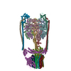
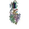
| Title | Thermus thermophilus V/A-type ATPase/synthase, rotational state 1R | ||||||
 Components Components |
| ||||||
 Keywords Keywords |  MEMBRANE PROTEIN / ATP hydrolysis/synthesis / proton translocation / rotary catalysis MEMBRANE PROTEIN / ATP hydrolysis/synthesis / proton translocation / rotary catalysis | ||||||
| Function / homology |  Function and homology information Function and homology informationproton-transporting V-type ATPase, V0 domain / proton-transporting two-sector ATPase complex, catalytic domain / proton-transporting ATP synthase complex / proton motive force-driven plasma membrane ATP synthesis / H+-transporting two-sector ATPase / proton-transporting ATPase activity, rotational mechanism / proton-transporting ATP synthase activity, rotational mechanism / ATP binding / metal ion bindingSimilarity search - Function | ||||||
| Biological species |   Thermus thermophilus (bacteria) Thermus thermophilus (bacteria) | ||||||
| Method | ELECTRON MICROSCOPY / single particle reconstruction / cryo EM / Resolution: 4.3 Å | ||||||
 Authors Authors | Zhou, L. / Sazanov, L. | ||||||
 Citation Citation | Journal: Science / Year: 2019 Title: Structure and conformational plasticity of the intact V/A-type ATPase. Authors: Long Zhou / Leonid A Sazanov /  Abstract: V (vacuolar)/A (archaeal)-type adenosine triphosphatases (ATPases), found in archaea and eubacteria, couple ATP hydrolysis or synthesis to proton translocation across the plasma membrane using the ...V (vacuolar)/A (archaeal)-type adenosine triphosphatases (ATPases), found in archaea and eubacteria, couple ATP hydrolysis or synthesis to proton translocation across the plasma membrane using the rotary-catalysis mechanism. They belong to the V-type ATPase family, which differs from the mitochondrial/chloroplast F-type ATP synthases in overall architecture. We solved cryo-electron microscopy structures of the intact V/A-ATPase, reconstituted into lipid nanodiscs, in three rotational states and two substates. These structures indicate substantial flexibility between V and V in a working enzyme, which results from mechanical competition between central shaft rotation and resistance from the peripheral stalks. We also describe details of adenosine diphosphate inhibition release, V-V torque transmission, and proton translocation, which are relevant for the entire V-type ATPase family. | ||||||
| History |
|
- Structure visualization
| Movie |
Movie viewer |
|---|---|
| Structure viewer | Molecule: MolmilJmol/JSmol |
- Downloads & links
-Download
| PDBx/mmCIF format | 6r10.cif.gz | 1008.9 KB | Display | PDBx/mmCIF format |
|---|---|---|---|---|
| PDB format | pdb6r10.ent.gz | 789 KB | Display | PDB format |
| PDBx/mmJSON format | 6r10.json.gz | Tree view | PDBx/mmJSON format | |
| Others |  Other downloads Other downloads |
-Validation report
| Arichive directory | https://data.pdbj.org/pub/pdb/validation_reports/r1/6r10ftp://data.pdbj.org/pub/pdb/validation_reports/r1/6r10 | HTTPS FTP |
|---|
-Related structure data
| Related structure data |  4703MC  4640C  4699C 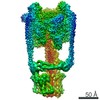 4700C  4702C  6qumC  6r0wC  6r0yC  6r0zC C: citing same article ( M: map data used to model this data |
|---|---|
| Similar structure data |


-Links
 PDBj
PDBj


- Assembly
| Deposited unit | 
|
|---|---|
| 1 |
|
-Components
-V-type ATP synthase ... , 7 types, 12 molecules ABCDEFGHJLMN
| #1: Protein | Mass: 63699.980 Da / Num. of mol.: 3 / Source method: isolated from a natural source Source: (natural) Thermus thermophilus (strain HB8 / ATCC 27634 / DSM 579) (bacteria)References: UniProt: Q56403, H+-transporting two-sector ATPase#2: Protein | Mass: 53219.500 Da / Num. of mol.: 3 / Source method: isolated from a natural source Source: (natural) Thermus thermophilus (strain HB8 / ATCC 27634 / DSM 579) (bacteria)References: UniProt: Q56404 #3: Protein | | Mass: 24715.566 Da / Num. of mol.: 1 / Source method: isolated from a natural source Source: (natural) Thermus thermophilus (strain HB8 / ATCC 27634 / DSM 579) (bacteria)References: UniProt: O87880 #4: Protein | | Mass: 11294.904 Da / Num. of mol.: 1 / Source method: isolated from a natural source Source: (natural) Thermus thermophilus (strain HB8 / ATCC 27634 / DSM 579) (bacteria)References: UniProt: P74903 #6: Protein | Mass: 20645.582 Da / Num. of mol.: 2 / Source method: isolated from a natural source Source: (natural) Thermus thermophilus (strain HB8 / ATCC 27634 / DSM 579) (bacteria)References: UniProt: P74901 #7: Protein | | Mass: 35968.570 Da / Num. of mol.: 1 / Source method: isolated from a natural source Source: (natural) Thermus thermophilus (strain HB8 / ATCC 27634 / DSM 579) (bacteria)References: UniProt: P74902 #8: Protein | | Mass: 72204.289 Da / Num. of mol.: 1 / Source method: isolated from a natural source Source: (natural) Thermus thermophilus (strain HB8 / ATCC 27634 / DSM 579) (bacteria)References: UniProt: Q5SIT6 |
|---|
-V-type ATP synthase, subunit ... , 2 types, 14 molecules IKOPQRSTUVWXYZ
| #5: Protein | Mass: 13166.218 Da / Num. of mol.: 2 / Source method: isolated from a natural source Source: (natural) Thermus thermophilus (strain HB8 / ATCC 27634 / DSM 579) (bacteria)References: UniProt: Q5SIT5 #9: Protein | Mass: 9841.714 Da / Num. of mol.: 12 / Source method: isolated from a natural source Source: (natural) Thermus thermophilus (strain HB8 / ATCC 27634 / DSM 579) (bacteria)References: UniProt: Q5SIT7 |
|---|
-Non-polymers , 2 types, 3 molecules 

| #10: Chemical | ChemComp-MG /  Mass: 24.305 Da / Num. of mol.: 1 / Source method: obtained synthetically / Formula: Mg Mass: 24.305 Da / Num. of mol.: 1 / Source method: obtained synthetically / Formula: Mg |
|---|---|
| #11: Chemical | Adenosine diphosphate Mass: 427.201 Da / Num. of mol.: 2 / Source method: obtained synthetically / Formula: C10H15N5O10P2 / Comment: ADP, energy-carrying molecule*YM Mass: 427.201 Da / Num. of mol.: 2 / Source method: obtained synthetically / Formula: C10H15N5O10P2 / Comment: ADP, energy-carrying molecule*YM |
-Experimental details
-Experiment
| Experiment | Method: ELECTRON MICROSCOPY |
|---|---|
| EM experiment | Aggregation state: PARTICLE / 3D reconstruction method: single particle reconstruction |
- Sample preparation
| Component | Name: Thermus thermophilus V/A-type ATPase/ATP synthase / Type: COMPLEX / Entity ID: #1-#9 / Source: NATURAL | ||||||||||||||||||||||||||||||||
|---|---|---|---|---|---|---|---|---|---|---|---|---|---|---|---|---|---|---|---|---|---|---|---|---|---|---|---|---|---|---|---|---|---|
| Molecular weight |
| ||||||||||||||||||||||||||||||||
| Source (natural) | Organism: Thermus thermophilus HB8 (bacteria) | ||||||||||||||||||||||||||||||||
| Buffer solution | pH: 8 | ||||||||||||||||||||||||||||||||
| Buffer component |
| ||||||||||||||||||||||||||||||||
| Specimen | Conc.: 5.4 mg/ml / Embedding applied: NO / Shadowing applied: NO / Staining applied: NO / Vitrification applied: YES | ||||||||||||||||||||||||||||||||
| Specimen support | Grid material: COPPER / Grid mesh size: 300 divisions/in. / Grid type: Quantifoil R0.6/1 | ||||||||||||||||||||||||||||||||
| Vitrification | Instrument: FEI VITROBOT MARK IV / Cryogen name: ETHANE / Humidity: 100 % / Chamber temperature: 277.15 K |
- Electron microscopy imaging
| Experimental equipment |  Model: Titan Krios / Image courtesy: FEI Company |
|---|---|
| Microscopy | Model: FEI TITAN KRIOS |
| Electron gun | Electron source: FIELD EMISSION GUN / Accelerating voltage: 300 kV / Illumination mode: SPOT SCAN |
| Electron lens | Mode: BRIGHT FIELDBright-field microscopy / Nominal magnification: 75000 X / Calibrated magnification: 129032 X / Nominal defocus max: 2000 nm / Nominal defocus min: 1000 nm / Calibrated defocus min: 500 nm / Calibrated defocus max: 1780 nm |
| Specimen holder | Cryogen: NITROGEN / Specimen holder model: FEI TITAN KRIOS AUTOGRID HOLDER |
| Image recording | Average exposure time: 63 sec. / Electron dose: 50 e/Å2 / Detector mode: COUNTING / Film or detector model: FEI FALCON III (4k x 4k) / Num. of grids imaged: 1 / Num. of real images: 2700 |
| Image scans | Sampling size: 14 µm / Width: 4096 / Height: 4096 |
- Processing
| Software | Name: PHENIX / Version: 1.13_2998: / Classification: refinement | ||||||||||||||||||||||||||||||||||||||||||||||||||||||||||||||||||||||||||||||||||||||||||||||||||||||||||||||||||||||||||||||||||||||||||||||||||||||||
|---|---|---|---|---|---|---|---|---|---|---|---|---|---|---|---|---|---|---|---|---|---|---|---|---|---|---|---|---|---|---|---|---|---|---|---|---|---|---|---|---|---|---|---|---|---|---|---|---|---|---|---|---|---|---|---|---|---|---|---|---|---|---|---|---|---|---|---|---|---|---|---|---|---|---|---|---|---|---|---|---|---|---|---|---|---|---|---|---|---|---|---|---|---|---|---|---|---|---|---|---|---|---|---|---|---|---|---|---|---|---|---|---|---|---|---|---|---|---|---|---|---|---|---|---|---|---|---|---|---|---|---|---|---|---|---|---|---|---|---|---|---|---|---|---|---|---|---|---|---|---|---|---|---|
| EM software |
| ||||||||||||||||||||||||||||||||||||||||||||||||||||||||||||||||||||||||||||||||||||||||||||||||||||||||||||||||||||||||||||||||||||||||||||||||||||||||
| CTF correction | Type: PHASE FLIPPING AND AMPLITUDE CORRECTION | ||||||||||||||||||||||||||||||||||||||||||||||||||||||||||||||||||||||||||||||||||||||||||||||||||||||||||||||||||||||||||||||||||||||||||||||||||||||||
| Particle selection | Num. of particles selected: 181245 | ||||||||||||||||||||||||||||||||||||||||||||||||||||||||||||||||||||||||||||||||||||||||||||||||||||||||||||||||||||||||||||||||||||||||||||||||||||||||
| Symmetry | Point symmetry: C1 (asymmetric) | ||||||||||||||||||||||||||||||||||||||||||||||||||||||||||||||||||||||||||||||||||||||||||||||||||||||||||||||||||||||||||||||||||||||||||||||||||||||||
| 3D reconstruction | Resolution: 4.3 Å / Resolution method: FSC 0.143 CUT-OFF / Num. of particles: 15571 / Algorithm: FOURIER SPACE / Num. of class averages: 1 / Symmetry type: POINT | ||||||||||||||||||||||||||||||||||||||||||||||||||||||||||||||||||||||||||||||||||||||||||||||||||||||||||||||||||||||||||||||||||||||||||||||||||||||||
| Atomic model building | B value: 167.6 / Space: REAL | ||||||||||||||||||||||||||||||||||||||||||||||||||||||||||||||||||||||||||||||||||||||||||||||||||||||||||||||||||||||||||||||||||||||||||||||||||||||||
| Atomic model building | 3D fitting-ID: 1 / Source name: PDB / Type: experimental model
|

