 Movie
Movie Controller

[English] 日本語
 Yorodumi
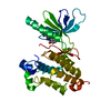
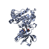
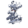
- PDB-6qvv: Structure and function of phenuiviridae cap snatching endonucleases -
+ Open data

- Basic information
| Entry | Database: PDB / ID: 6qvv | ||||||
|---|---|---|---|---|---|---|---|
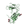
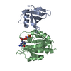
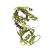
| Title | Structure and function of phenuiviridae cap snatching endonucleases | ||||||
 Components Components | RNA-dependent RNA polymerase | ||||||
 Keywords Keywords | HYDROLASE / Cap snatching endonuclease of Toscana virus at the N-term of the L protein. | ||||||
| Function / homology |  Function and homology information Function and homology informationRNA-templated viral transcription / nucleoside binding / negative stranded viral RNA replication / host cell endoplasmic reticulum / endoplasmic reticulum-Golgi intermediate compartment / virion component / host cell endoplasmic reticulum-Golgi intermediate compartment / host cell Golgi apparatus / Hydrolases; Acting on ester bonds / hydrolase activity ...RNA-templated viral transcription / nucleoside binding / negative stranded viral RNA replication / host cell endoplasmic reticulum / endoplasmic reticulum-Golgi intermediate compartment / virion component / host cell endoplasmic reticulum-Golgi intermediate compartment / host cell Golgi apparatus / Hydrolases; Acting on ester bonds / hydrolase activity / RNA-directed RNA polymerase / viral RNA genome replication / RNA-dependent RNA polymerase activity / nucleotide binding / DNA-templated transcription / metal ion bindingSimilarity search - Function | ||||||
| Biological species |  Toscana virus Toscana virus | ||||||
| Method | X-RAY DIFFRACTION / SYNCHROTRON / MOLECULAR REPLACEMENT / Resolution: 2.4 Å | ||||||
 Authors Authors | Reguera, J. / Jones, R. / Bragagniolo, G. / Lessoued, S. / Mate, M. | ||||||
| Funding support |  France, 1items France, 1items
| ||||||
 Citation Citation | Journal: Nucleic Acids Res. / Year: 2019 Title: Structure and function of the Toscana virus cap-snatching endonuclease. Authors: Jones, R. / Lessoued, S. / Meier, K. / Devignot, S. / Barata-Garcia, S. / Mate, M. / Bragagnolo, G. / Weber, F. / Rosenthal, M. / Reguera, J. | ||||||
| History |
|
- Structure visualization
| Structure viewer | Molecule: MolmilJmol/JSmol |
|---|
- Downloads & links
-Download
| PDBx/mmCIF format | 6qvv.cif.gz | 95.9 KB | Display | PDBx/mmCIF format |
|---|---|---|---|---|
| PDB format | pdb6qvv.ent.gz | 72.5 KB | Display | PDB format |
| PDBx/mmJSON format | 6qvv.json.gz | Tree view | PDBx/mmJSON format | |
| Others |  Other downloads Other downloads |
-Validation report
| Arichive directory | https://data.pdbj.org/pub/pdb/validation_reports/qv/6qvvftp://data.pdbj.org/pub/pdb/validation_reports/qv/6qvv | HTTPS FTP |
|---|
-Related structure data
| Related structure data |  6qw0C 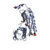 6qw5SC S: Starting model for refinement C: citing same article ( |
|---|---|
| Similar structure data |


-Links
 PDBj
PDBj
- Assembly
| Deposited unit | 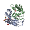
| ||||||||
|---|---|---|---|---|---|---|---|---|---|
| 1 | 
| ||||||||
| 2 | 
| ||||||||
| Unit cell |
|
-Components
| #1: Protein | / cap-snatching endonuclease Mass: 23751.697 Da / Num. of mol.: 2 Source method: isolated from a genetically manipulated source Source: (gene. exp.) Toscana virus / Production host:  Escherichia coli BL21(DE3) (bacteria) / Variant (production host): Gold pLysS AG / References: UniProt: S4ZA26, UniProt: P37800*PLUS Escherichia coli BL21(DE3) (bacteria) / Variant (production host): Gold pLysS AG / References: UniProt: S4ZA26, UniProt: P37800*PLUS#2: Chemical | Manganese  Mass: 54.938 Da / Num. of mol.: 2 / Source method: obtained synthetically / Formula: Mn Mass: 54.938 Da / Num. of mol.: 2 / Source method: obtained synthetically / Formula: Mn#3: Chemical | ChemComp-SO4 / Sulfate  Mass: 96.063 Da / Num. of mol.: 10 / Source method: obtained synthetically / Formula: SO4 Mass: 96.063 Da / Num. of mol.: 10 / Source method: obtained synthetically / Formula: SO4#4: Chemical | ChemComp-GOL / | Glycerol  Mass: 92.094 Da / Num. of mol.: 1 / Source method: obtained synthetically / Formula: C3H8O3 Mass: 92.094 Da / Num. of mol.: 1 / Source method: obtained synthetically / Formula: C3H8O3#5: Water | ChemComp-HOH / | Water Mass: 18.015 Da / Num. of mol.: 132 / Source method: isolated from a natural source / Formula: H2O Mass: 18.015 Da / Num. of mol.: 132 / Source method: isolated from a natural source / Formula: H2O |
|---|
-Experimental details
-Experiment
| Experiment | Method: X-RAY DIFFRACTION / Number of used crystals: 1 |
|---|
- Sample preparation
| Crystal | Density Matthews: 3.41 Å3/Da / Density % sol: 63.91 % |
|---|---|
| Crystal grow | Temperature: 293 K / Method: counter-diffusion / Details: 0.1 M Hepes pH 7.5 0.75 M LiSO4 |
-Data collection
| Diffraction | Mean temperature: 100 K / Serial crystal experiment: N |
|---|---|
| Diffraction source | Source: SYNCHROTRON / Site: ESRF / Beamline: MASSIF-1 / Wavelength: 0.9677 Å |
| Detector | Type: DECTRIS EIGER R 1M / Detector: PIXEL / Date: Jul 22, 2017 |
| Radiation | Protocol: SINGLE WAVELENGTH / Monochromatic (M) / Laue (L): M / Scattering type: x-ray |
| Radiation wavelength | Wavelength: 0.9677 Å / Relative weight: 1 |
| Reflection | Resolution: 2.4→48.18 Å / Num. obs: 25558 / % possible obs: 98.7 % / Redundancy: 4.2 % / CC1/2: 0.993 / Rmerge(I) obs: 0.159 / Rpim(I) all: 0.086 / Rrim(I) all: 0.182 / Χ2: 0.99 / Net I/σ(I): 7.3 |
| Reflection shell | Resolution: 2.4→2.49 Å / Redundancy: 4.4 % / Rmerge(I) obs: 1.062 / Mean I/σ(I) obs: 1.4 / Num. unique obs: 2672 / CC1/2: 0.593 / Rpim(I) all: 0.57 / Rrim(I) all: 1.211 / Χ2: 0.98 / % possible all: 99.1 |
- Processing
| Software |
| ||||||||||||||||||||||||||||||||||||||||||||||||||||||||||||||||||||||
|---|---|---|---|---|---|---|---|---|---|---|---|---|---|---|---|---|---|---|---|---|---|---|---|---|---|---|---|---|---|---|---|---|---|---|---|---|---|---|---|---|---|---|---|---|---|---|---|---|---|---|---|---|---|---|---|---|---|---|---|---|---|---|---|---|---|---|---|---|---|---|---|
| Refinement | Method to determine structure: MOLECULAR REPLACEMENT Starting model: 6QW5 Resolution: 2.4→48.18 Å / SU ML: 0.32 / Cross valid method: FREE R-VALUE / σ(F): 1.34 / Phase error: 26.34 / Stereochemistry target values: ML
| ||||||||||||||||||||||||||||||||||||||||||||||||||||||||||||||||||||||
| Solvent computation | Shrinkage radii: 0.9 Å / VDW probe radii: 1.11 Å / Solvent model: FLAT BULK SOLVENT MODEL | ||||||||||||||||||||||||||||||||||||||||||||||||||||||||||||||||||||||
| Refinement step | Cycle: LAST / Resolution: 2.4→48.18 Å
| ||||||||||||||||||||||||||||||||||||||||||||||||||||||||||||||||||||||
| Refine LS restraints |
| ||||||||||||||||||||||||||||||||||||||||||||||||||||||||||||||||||||||
| LS refinement shell |
|
