 Movie
Movie Controller


+ Open data

- Basic information
| Entry | Database: PDB / ID: 6qcc | ||||||||||||
|---|---|---|---|---|---|---|---|---|---|---|---|---|---|
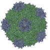
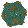
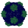
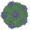
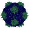
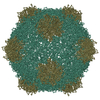
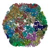
| Title | Cryo-EM Atomic Structure of Broad Bean Stain Virus (BBSV) | ||||||||||||
 Components Components |
| ||||||||||||
 Keywords Keywords |  VIRUS / comovirus capsid plant virus BBSV VIRUS / comovirus capsid plant virus BBSV | ||||||||||||
| Function / homology |  Function and homology information Function and homology informationtransport of virus in host, cell to cell / host cell plasmodesma / T=3 icosahedral viral capsid / membrane => GO:0016020 / GTP binding / structural molecule activity / DNA binding / RNA bindingSimilarity search - Function | ||||||||||||
| Biological species |  Broad bean stain virus Broad bean stain virus | ||||||||||||
| Method | ELECTRON MICROSCOPY / single particle reconstruction / cryo EM / Resolution: 3.22 Å | ||||||||||||
 Authors Authors | Lecorre, F. / Lai Jee Him, J. / Blanc, S. / Zeddam, J.-L. / Trapani, S. / Bron, P. | ||||||||||||
| Funding support | New Caledonia,  France, 3items France, 3items
| ||||||||||||
 Citation Citation | Journal: Virology / Year: 2019 Title: The cryo-electron microscopy structure of Broad Bean Stain Virus suggests a common capsid assembly mechanism among comoviruses. Authors: François Lecorre / Joséphine Lai-Kee-Him / Stéphane Blanc / Jean-Louis Zeddam / Stefano Trapani / Patrick Bron / Abstract: The Broad bean stain virus (BBSV) is a member of the genus Comovirus infecting Fabaceae. The virus is transmitted through seed and by plant weevils causing severe and widespread disease worldwide. ...The Broad bean stain virus (BBSV) is a member of the genus Comovirus infecting Fabaceae. The virus is transmitted through seed and by plant weevils causing severe and widespread disease worldwide. BBSV has a bipartite, positive-sense, single-stranded RNA genome encapsidated in icosahedral particles. We present here the cryo-electron microscopy reconstruction of the BBSV and an atomic model of the capsid proteins refined at 3.22 Å resolution. We identified residues involved in RNA/capsid interactions revealing a unique RNA genome organization. Inspection of the small coat protein C-terminal domain highlights a maturation cleavage between Leu567 and Leu568 and interactions of the C-terminal stretch with neighbouring small coat proteins within the capsid pentameric turrets. These interactions previously proposed to play a key role in the assembly of the Cowpea mosaic virus suggest a common capsid assembly mechanism throughout all comovirus species. | ||||||||||||
| History |
|
- Structure visualization
| Movie |
Movie viewer |
|---|---|
| Structure viewer | Molecule: MolmilJmol/JSmol |
- Downloads & links
-Download
| PDBx/mmCIF format | 6qcc.cif.gz | 106.8 KB | Display | PDBx/mmCIF format |
|---|---|---|---|---|
| PDB format | pdb6qcc.ent.gz | 81.1 KB | Display | PDB format |
| PDBx/mmJSON format | 6qcc.json.gz | Tree view | PDBx/mmJSON format | |
| Others |  Other downloads Other downloads |
-Validation report
| Arichive directory | https://data.pdbj.org/pub/pdb/validation_reports/qc/6qccftp://data.pdbj.org/pub/pdb/validation_reports/qc/6qcc | HTTPS FTP |
|---|
-Related structure data
| Related structure data | 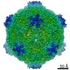 4504MC M: map data used to model this data C: citing same article ( |
|---|---|
| Similar structure data |


-Links
 PDBj
PDBj
- Assembly
| Deposited unit | 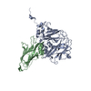
|
|---|---|
| 1 | x 60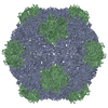
|
| 2 |
|
| 3 | x 5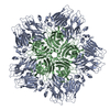
|
| 4 | x 6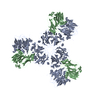
|
| 5 | 
|
| Symmetry | Point symmetry: (Schoenflies symbol: I (icosahedral)) |
-Components
| #1: Protein | Mass: 41247.977 Da / Num. of mol.: 1 / Source method: isolated from a natural source / Source: (natural) Broad bean stain virus / References: UniProt: D0PSV7 |
|---|---|
| #2: Protein | Mass: 23589.662 Da / Num. of mol.: 1 / Source method: isolated from a natural source / Source: (natural) Broad bean stain virus / References: UniProt: D0PSV7 |
-Experimental details
-Experiment
| Experiment | Method: ELECTRON MICROSCOPY |
|---|---|
| EM experiment | Aggregation state: PARTICLE / 3D reconstruction method: single particle reconstruction |
- Sample preparation
| Component | Name: Broad bean stain virus / Type: VIRUS / Entity ID: all / Source: NATURAL |
|---|---|
| Molecular weight | Experimental value: NO |
| Source (natural) | Organism: Broad bean stain virus |
| Details of virus | Empty: NO / Enveloped: NO / Isolate: STRAIN / Type: VIRION |
| Natural host | Organism: Vicia faba |
| Virus shell | Name: capsid / Diameter: 300 nm / Triangulation number (T number): 1 |
| Buffer solution | pH: 7.4 |
| Buffer component | Conc.: 20 mM / Name: Phosphate / Formula: K2HPO4/KH2PO4 |
| Specimen | Conc.: 2 mg/ml / Embedding applied: NO / Shadowing applied: NO / Staining applied: NO / Vitrification applied: YES |
| Vitrification | Instrument: GATAN CRYOPLUNGE 3 / Cryogen name: ETHANE / Humidity: 99 % / Chamber temperature: 298.15 K / Details: blot for 1 second before plunging |
- Electron microscopy imaging
| Experimental equipment |  Model: Titan Krios / Image courtesy: FEI Company |
|---|---|
| Microscopy | Model: FEI TITAN KRIOS |
| Electron gun | Electron source: FIELD EMISSION GUN / Accelerating voltage: 300 kV / Illumination mode: FLOOD BEAM |
| Electron lens | Mode: BRIGHT FIELDBright-field microscopy / Nominal magnification: 59000 X |
| Specimen holder | Cryogen: NITROGEN / Specimen holder model: FEI TITAN KRIOS AUTOGRID HOLDER |
| Image recording | Average exposure time: 2 sec. / Electron dose: 45 e/Å2 / Detector mode: INTEGRATING / Film or detector model: FEI FALCON II (4k x 4k) / Num. of grids imaged: 1 / Num. of real images: 2899 / Details: 1494 images were retained for 3D reconstruction |
| Image scans | Sampling size: 14 µm / Width: 2048 / Height: 2048 / Movie frames/image: 35 / Used frames/image: 3-9 |
- Processing
| EM software |
| |||||||||||||||||||||||||||||||||||||||||||||||||||||||||||||||||
|---|---|---|---|---|---|---|---|---|---|---|---|---|---|---|---|---|---|---|---|---|---|---|---|---|---|---|---|---|---|---|---|---|---|---|---|---|---|---|---|---|---|---|---|---|---|---|---|---|---|---|---|---|---|---|---|---|---|---|---|---|---|---|---|---|---|---|
| CTF correction | Type: PHASE FLIPPING AND AMPLITUDE CORRECTION | |||||||||||||||||||||||||||||||||||||||||||||||||||||||||||||||||
| Particle selection | Num. of particles selected: 35663 | |||||||||||||||||||||||||||||||||||||||||||||||||||||||||||||||||
| Symmetry | Point symmetry: I (icosahedral) | |||||||||||||||||||||||||||||||||||||||||||||||||||||||||||||||||
| 3D reconstruction | Resolution: 3.22 Å / Resolution method: FSC 0.143 CUT-OFF / Num. of particles: 17233 / Algorithm: FOURIER SPACE / Num. of class averages: 1 / Symmetry type: POINT | |||||||||||||||||||||||||||||||||||||||||||||||||||||||||||||||||
| Atomic model building | Protocol: AB INITIO MODEL / Space: REAL | |||||||||||||||||||||||||||||||||||||||||||||||||||||||||||||||||
| Atomic model building | PDB-ID: 1NY7 Accession code: 1NY7 / Source name: PDB / Type: experimental model |

