 Movie
Movie Controller


+ Open data

- Basic information
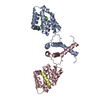
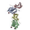
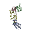
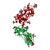
| Entry | Database: PDB / ID: 6q8j | ||||||
|---|---|---|---|---|---|---|---|
| Title | Nterminal domain of human SMU1 in complex with LSP641 | ||||||
 Components Components | WD40 repeat-containing protein SMU1 | ||||||
 Keywords Keywords |  SPLICING / Splicing factor / influenza virus SPLICING / Splicing factor / influenza virus | ||||||
| Function / homology |  Function and homology information Function and homology informationU2-type precatalytic spliceosome / precatalytic spliceosome / regulation of alternative mRNA splicing, via spliceosome / mRNA Splicing - Major Pathway / RNA splicing / mRNA splicing, via spliceosome / nuclear speck / nucleoplasm / nucleus / cytoplasmSimilarity search - Function | ||||||
| Biological species |  Homo sapiens (human) Homo sapiens (human) | ||||||
| Method | X-RAY DIFFRACTION / SYNCHROTRON / MOLECULAR REPLACEMENT / Resolution: 1.8 Å | ||||||
 Authors Authors | Tengo, L. / Le Corre, L. / Fournier, G. / Ashraf, U. / Busca, P. / Rameix-Welti, M.-A. / Gravier-Pelletier, C. / Ruigrok, R.W.H. / Jacob, Y. / Vidalain, P.-O. ...Tengo, L. / Le Corre, L. / Fournier, G. / Ashraf, U. / Busca, P. / Rameix-Welti, M.-A. / Gravier-Pelletier, C. / Ruigrok, R.W.H. / Jacob, Y. / Vidalain, P.-O. / Pietrancosta, N. / Naffakh, N. / McCarthy, A.A. / Crepin, T. | ||||||
 Citation Citation | Journal: Proc.Natl.Acad.Sci.USA / Year: 2019 Title: Destabilization of the human RED-SMU1 splicing complex as a basis for host-directed antiinfluenza strategy. Authors: Ashraf, U. / Tengo, L. / Le Corre, L. / Fournier, G. / Busca, P. / McCarthy, A.A. / Rameix-Welti, M.A. / Gravier-Pelletier, C. / Ruigrok, R.W.H. / Jacob, Y. / Vidalain, P.O. / Pietrancosta, ...Authors: Ashraf, U. / Tengo, L. / Le Corre, L. / Fournier, G. / Busca, P. / McCarthy, A.A. / Rameix-Welti, M.A. / Gravier-Pelletier, C. / Ruigrok, R.W.H. / Jacob, Y. / Vidalain, P.O. / Pietrancosta, N. / Crepin, T. / Naffakh, N. | ||||||
| History |
|
- Structure visualization
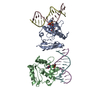
| Structure viewer | Molecule: MolmilJmol/JSmol |
|---|
- Downloads & links
-Download
| PDBx/mmCIF format | 6q8j.cif.gz | 101.8 KB | Display | PDBx/mmCIF format |
|---|---|---|---|---|
| PDB format | pdb6q8j.ent.gz | 77.3 KB | Display | PDB format |
| PDBx/mmJSON format | 6q8j.json.gz | Tree view | PDBx/mmJSON format | |
| Others |  Other downloads Other downloads |
-Validation report
| Arichive directory | https://data.pdbj.org/pub/pdb/validation_reports/q8/6q8jftp://data.pdbj.org/pub/pdb/validation_reports/q8/6q8j | HTTPS FTP |
|---|
-Related structure data
| Related structure data |  6q8fSC  6q8iC S: Starting model for refinement C: citing same article ( |
|---|---|
| Similar structure data |


-Links
 PDBj
PDBj

- Assembly
| Deposited unit | 
| ||||||||
|---|---|---|---|---|---|---|---|---|---|
| 1 | 
| ||||||||
| Unit cell |
|
-Components
| #1: Protein | Mass: 22115.436 Da / Num. of mol.: 1 Source method: isolated from a genetically manipulated source Source: (gene. exp.) Homo sapiens (human) / Gene: SMU1 / Production host:  Escherichia coli (E. coli) / References: UniProt: Q2TAY7 Escherichia coli (E. coli) / References: UniProt: Q2TAY7 | ||
|---|---|---|---|
| #2: Chemical |   Mass: 650.773 Da / Num. of mol.: 2 / Source method: obtained synthetically / Formula: C35H42N10O3 Mass: 650.773 Da / Num. of mol.: 2 / Source method: obtained synthetically / Formula: C35H42N10O3#3: Water | ChemComp-HOH / | Water Mass: 18.015 Da / Num. of mol.: 102 / Source method: isolated from a natural source / Formula: H2O Mass: 18.015 Da / Num. of mol.: 102 / Source method: isolated from a natural source / Formula: H2O |
-Experimental details
-Experiment
| Experiment | Method: X-RAY DIFFRACTION / Number of used crystals: 1 |
|---|
- Sample preparation
| Crystal | Density Matthews: 3.12 Å3/Da / Density % sol: 60.52 % |
|---|---|
| Crystal grow | Temperature: 293 K / Method: vapor diffusion, sitting drop / pH: 6 Details: 0.1 M Bis-Tris pH 6, 16-20 % PEG 10K, 0.2 M ammonium acetate |
-Data collection
| Diffraction | Mean temperature: 100 K / Serial crystal experiment: N |
|---|---|
| Diffraction source | Source: SYNCHROTRON / Site: ESRF  / Beamline: ID30B / Wavelength: 0.97625 Å / Beamline: ID30B / Wavelength: 0.97625 Å |
| Detector | Type: DECTRIS PILATUS3 6M / Detector: PIXEL / Date: Feb 25, 2016 |
| Radiation | Protocol: SINGLE WAVELENGTH / Monochromatic (M) / Laue (L): M / Scattering type: x-ray |
| Radiation wavelength | Wavelength: 0.97625 Å / Relative weight: 1 |
| Reflection | Resolution: 1.8→44 Å / Num. obs: 26383 / % possible obs: 97.4 % / Redundancy: 5.27 % / Rrim(I) all: 0.051 / Net I/σ(I): 16.86 |
| Reflection shell | Resolution: 1.8→1.88 Å / Redundancy: 5.32 % / Mean I/σ(I) obs: 2.2 / Num. unique obs: 3140 / Rrim(I) all: 0.762 / % possible all: 97.7 |
- Processing
| Software |
| ||||||||||||||||||||||||||||||||||||||||||||||||||||||||||||||||||||||||||||||||||||||||||||||||||||||||||||||||||||||||||||||||||||||||||||||||||||||||||||||||||||||||||||||||||||||
|---|---|---|---|---|---|---|---|---|---|---|---|---|---|---|---|---|---|---|---|---|---|---|---|---|---|---|---|---|---|---|---|---|---|---|---|---|---|---|---|---|---|---|---|---|---|---|---|---|---|---|---|---|---|---|---|---|---|---|---|---|---|---|---|---|---|---|---|---|---|---|---|---|---|---|---|---|---|---|---|---|---|---|---|---|---|---|---|---|---|---|---|---|---|---|---|---|---|---|---|---|---|---|---|---|---|---|---|---|---|---|---|---|---|---|---|---|---|---|---|---|---|---|---|---|---|---|---|---|---|---|---|---|---|---|---|---|---|---|---|---|---|---|---|---|---|---|---|---|---|---|---|---|---|---|---|---|---|---|---|---|---|---|---|---|---|---|---|---|---|---|---|---|---|---|---|---|---|---|---|---|---|---|---|
| Refinement | Method to determine structure: MOLECULAR REPLACEMENT Starting model: 6Q8F Resolution: 1.8→43.72 Å / Cor.coef. Fo:Fc: 0.971 / Cor.coef. Fo:Fc free: 0.957 / SU B: 6.684 / SU ML: 0.088 / Cross valid method: THROUGHOUT / ESU R: 0.125 / ESU R Free: 0.106 / Details: HYDROGENS HAVE BEEN ADDED IN THE RIDING POSITIONS
| ||||||||||||||||||||||||||||||||||||||||||||||||||||||||||||||||||||||||||||||||||||||||||||||||||||||||||||||||||||||||||||||||||||||||||||||||||||||||||||||||||||||||||||||||||||||
| Solvent computation | Ion probe radii: 0.8 Å / Shrinkage radii: 0.8 Å / VDW probe radii: 1.2 Å | ||||||||||||||||||||||||||||||||||||||||||||||||||||||||||||||||||||||||||||||||||||||||||||||||||||||||||||||||||||||||||||||||||||||||||||||||||||||||||||||||||||||||||||||||||||||
| Displacement parameters | Biso mean: 43.95 Å2
| ||||||||||||||||||||||||||||||||||||||||||||||||||||||||||||||||||||||||||||||||||||||||||||||||||||||||||||||||||||||||||||||||||||||||||||||||||||||||||||||||||||||||||||||||||||||
| Refinement step | Cycle: 1 / Resolution: 1.8→43.72 Å
| ||||||||||||||||||||||||||||||||||||||||||||||||||||||||||||||||||||||||||||||||||||||||||||||||||||||||||||||||||||||||||||||||||||||||||||||||||||||||||||||||||||||||||||||||||||||
| Refine LS restraints |
|