 Movie
Movie Controller

[English] 日本語
 Yorodumi
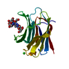
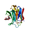
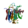
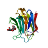
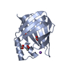
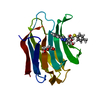
- PDB-6q17: Crystal structure of Human galectin-3 CRD in complex with Methyl ... -
+ Open data

- Basic information
| Entry | Database: PDB / ID: 6q17 | ||||||
|---|---|---|---|---|---|---|---|
| Title | Crystal structure of Human galectin-3 CRD in complex with Methyl 3-O-[1-(b-D-galactopyranosyl)-1,2,3-triazol-4-yl]-methyl-b-D-galactopyranoside | ||||||
 Components Components | Galectin-3 | ||||||
 Keywords Keywords | SUGAR BINDING PROTEIN | ||||||
| Function / homology |  Function and homology information Function and homology informationnegative regulation of protein tyrosine phosphatase activity / negative regulation of immunological synapse formation / negative regulation of T cell activation via T cell receptor contact with antigen bound to MHC molecule on antigen presenting cell / RUNX2 regulates genes involved in differentiation of myeloid cells / disaccharide binding / regulation of T cell apoptotic process / mononuclear cell migration / IgE binding / negative regulation of endocytosis / positive regulation of mononuclear cell migration ...negative regulation of protein tyrosine phosphatase activity / negative regulation of immunological synapse formation / negative regulation of T cell activation via T cell receptor contact with antigen bound to MHC molecule on antigen presenting cell / RUNX2 regulates genes involved in differentiation of myeloid cells / disaccharide binding / regulation of T cell apoptotic process / mononuclear cell migration / IgE binding / negative regulation of endocytosis / positive regulation of mononuclear cell migration / eosinophil chemotaxis / regulation of extrinsic apoptotic signaling pathway via death domain receptors / RUNX1 regulates transcription of genes involved in differentiation of myeloid cells / protein phosphatase inhibitor activity / negative regulation of T cell receptor signaling pathway / positive chemotaxis / regulation of T cell proliferation / positive regulation of calcium ion import / macrophage chemotaxis / chemoattractant activity / monocyte chemotaxis / Advanced glycosylation endproduct receptor signaling / ficolin-1-rich granule membrane / immunological synapse / laminin binding / epithelial cell differentiation / neutrophil chemotaxis / RNA splicing / secretory granule membrane / molecular condensate scaffold activity / negative regulation of extrinsic apoptotic signaling pathway / positive regulation of protein localization to plasma membrane / spliceosomal complex / positive regulation of protein-containing complex assembly / mRNA processing / carbohydrate binding / protein phosphatase binding / collagen-containing extracellular matrix / mitochondrial inner membrane / innate immune response / Neutrophil degranulation / cell surface / extracellular space / RNA binding / extracellular exosome / extracellular region / nucleoplasm / membrane / nucleus / plasma membrane / cytosol / cytoplasmSimilarity search - Function | ||||||
| Biological species |  Homo sapiens (human) Homo sapiens (human) | ||||||
| Method | X-RAY DIFFRACTION / SYNCHROTRON / MOLECULAR REPLACEMENT / Resolution: 1.98 Å | ||||||
 Authors Authors | Kishor, C. / Blanchard, H. | ||||||
| Funding support |  Australia, 1items Australia, 1items
| ||||||
 Citation Citation | Journal: Chem.Biol.Drug Des. / Year: 2020 Title: Linear triazole-linked pseudo oligogalactosides as scaffolds for galectin inhibitor development. Authors: Dussouy, C. / Kishor, C. / Lambert, A. / Lamoureux, C. / Blanchard, H. / Grandjean, C. | ||||||
| History |
|
- Structure visualization
| Structure viewer | Molecule: MolmilJmol/JSmol |
|---|
- Downloads & links
-Download
| PDBx/mmCIF format | 6q17.cif.gz | 55.3 KB | Display | PDBx/mmCIF format |
|---|---|---|---|---|
| PDB format | pdb6q17.ent.gz | 31 KB | Display | PDB format |
| PDBx/mmJSON format | 6q17.json.gz | Tree view | PDBx/mmJSON format | |
| Others |  Other downloads Other downloads |
-Validation report
| Arichive directory | https://data.pdbj.org/pub/pdb/validation_reports/q1/6q17ftp://data.pdbj.org/pub/pdb/validation_reports/q1/6q17 | HTTPS FTP |
|---|
-Related structure data


-Links
 PDBj
PDBj


- Assembly
| Deposited unit | 
| ||||||||||||
|---|---|---|---|---|---|---|---|---|---|---|---|---|---|
| 1 |
| ||||||||||||
| Unit cell |
|
-Components
| #1: Protein | / Gal-3 / 35 kDa lectin / Carbohydrate-binding protein 35 / CBP 35 / Galactose-specific lectin 3 / ...Gal-3 / 35 kDa lectin / Carbohydrate-binding protein 35 / CBP 35 / Galactose-specific lectin 3 / Galactoside-binding protein / GALBP / IgE-binding protein / L-31 / Laminin-binding protein / Lectin L-29 / Mac-2 antigen Mass: 15758.100 Da / Num. of mol.: 1 Source method: isolated from a genetically manipulated source Source: (gene. exp.) Homo sapiens (human) / Gene: LGALS3, MAC2 / Production host:  Escherichia coli (E. coli) / References: UniProt: P17931 Escherichia coli (E. coli) / References: UniProt: P17931 | ||||
|---|---|---|---|---|---|
| #2: Chemical | ChemComp-P8J /   Mass: 437.399 Da / Num. of mol.: 1 / Source method: isolated from a natural source / Formula: C16H27N3O11 / Feature type: SUBJECT OF INVESTIGATION Mass: 437.399 Da / Num. of mol.: 1 / Source method: isolated from a natural source / Formula: C16H27N3O11 / Feature type: SUBJECT OF INVESTIGATION | ||||
| #3: Chemical | Chloride  Mass: 35.453 Da / Num. of mol.: 2 / Source method: obtained synthetically / Formula: Cl Mass: 35.453 Da / Num. of mol.: 2 / Source method: obtained synthetically / Formula: Cl#4: Water | ChemComp-HOH / | Water Mass: 18.015 Da / Num. of mol.: 108 / Source method: isolated from a natural source / Formula: H2O Mass: 18.015 Da / Num. of mol.: 108 / Source method: isolated from a natural source / Formula: H2OHas ligand of interest | Y | |
-Experimental details
-Experiment
| Experiment | Method: X-RAY DIFFRACTION / Number of used crystals: 1 |
|---|
- Sample preparation
| Crystal | Density Matthews: 2.02 Å3/Da / Density % sol: 39.24 % |
|---|---|
| Crystal grow | Temperature: 297 K / Method: vapor diffusion, hanging drop / pH: 7.5 Details: 31% PEG 3100, 100 mM tris-HCl pH 7.5, 100 mM MgCl2, 8 mM 2-Mercaptoethanol |
-Data collection
| Diffraction | Mean temperature: 100 K / Serial crystal experiment: N |
|---|---|
| Diffraction source | Source: SYNCHROTRON / Site: Australian Synchrotron / Beamline: MX1 / Wavelength: 0.9537 Å |
| Detector | Type: ADSC QUANTUM 210 / Detector: CCD / Date: Mar 9, 2017 |
| Radiation | Protocol: SINGLE WAVELENGTH / Monochromatic (M) / Laue (L): M / Scattering type: x-ray |
| Radiation wavelength | Wavelength: 0.9537 Å / Relative weight: 1 |
| Reflection | Resolution: 1.98→42.48 Å / Num. obs: 9412 / % possible obs: 100 % / Redundancy: 11.5 % / Biso Wilson estimate: 11.613760561 Å2 / Rmerge(I) obs: 0.067 / Net I/σ(I): 27 |
| Reflection shell | Resolution: 1.99→2.06 Å / Rmerge(I) obs: 0.11 / Num. unique obs: 906 |
- Processing
| Software |
| ||||||||||||||||||||||||||||
|---|---|---|---|---|---|---|---|---|---|---|---|---|---|---|---|---|---|---|---|---|---|---|---|---|---|---|---|---|---|
| Refinement | Method to determine structure: MOLECULAR REPLACEMENT / Resolution: 1.98→42.4712432161 Å / SU ML: 0.183560541566 / Cross valid method: FREE R-VALUE / σ(F): 1.35131633093 / Phase error: 20.7599755456
| ||||||||||||||||||||||||||||
| Solvent computation | Shrinkage radii: 0.9 Å / VDW probe radii: 1.11 Å | ||||||||||||||||||||||||||||
| Displacement parameters | Biso mean: 12.949859417 Å2 | ||||||||||||||||||||||||||||
| Refinement step | Cycle: LAST / Resolution: 1.98→42.4712432161 Å
| ||||||||||||||||||||||||||||
| Refine LS restraints |
| ||||||||||||||||||||||||||||
| LS refinement shell |
|
