 ムービー
ムービー コントローラー


+ データを開く

- 基本情報
基本情報
| 登録情報 | データベース: PDB / ID: 6ptl | |||||||||
|---|---|---|---|---|---|---|---|---|---|---|
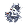
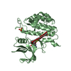
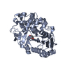
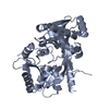
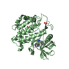
| タイトル | Structure of the self-association domain of the chromatin looping factor LDB1 | |||||||||
 要素 要素 | LIM domain-binding protein 1 | |||||||||
 キーワード キーワード |  GENE REGULATION (遺伝子発現の調節) / Dimer / Chromatin looping factor / nuclear protein / transcriptional regulator GENE REGULATION (遺伝子発現の調節) / Dimer / Chromatin looping factor / nuclear protein / transcriptional regulator | |||||||||
| 機能・相同性 |  機能・相同性情報regulation of kinase activity / cellular component assembly / negative regulation of erythrocyte differentiation / positive regulation of hemoglobin biosynthetic process / cerebellar Purkinje cell differentiation / epithelial structure maintenance / head development / primitive erythrocyte differentiation / beta-catenin-TCF complex / transcription-dependent tethering of RNA polymerase II gene DNA at nuclear periphery ...regulation of kinase activity / cellular component assembly / negative regulation of erythrocyte differentiation / positive regulation of hemoglobin biosynthetic process / cerebellar Purkinje cell differentiation / epithelial structure maintenance / head development / primitive erythrocyte differentiation / beta-catenin-TCF complex / transcription-dependent tethering of RNA polymerase II gene DNA at nuclear periphery / RUNX1 regulates transcription of genes involved in differentiation of HSCs / LIM domain binding / gastrulation with mouth forming second / anterior/posterior axis specification / regulation of focal adhesion assembly / cell leading edge / somatic stem cell population maintenance / positive regulation of cell adhesion / hair follicle development / regulation of cell migration / cerebellum development / positive regulation of transcription elongation by RNA polymerase II / neuron differentiation / Wntシグナル経路 / RNA polymerase II transcription regulator complex / : / nervous system development / DNA-binding transcription factor binding / RNA polymerase II-specific DNA-binding transcription factor binding / transcription regulator complex / transcription by RNA polymerase II / transcription coactivator activity / 細胞接着 / chromatin binding / クロマチン / enzyme binding / negative regulation of transcription by RNA polymerase II / protein homodimerization activity / positive regulation of transcription by RNA polymerase II / protein-containing complex / 核質 / identical protein binding / 細胞核 機能・相同性情報regulation of kinase activity / cellular component assembly / negative regulation of erythrocyte differentiation / positive regulation of hemoglobin biosynthetic process / cerebellar Purkinje cell differentiation / epithelial structure maintenance / head development / primitive erythrocyte differentiation / beta-catenin-TCF complex / transcription-dependent tethering of RNA polymerase II gene DNA at nuclear periphery ...regulation of kinase activity / cellular component assembly / negative regulation of erythrocyte differentiation / positive regulation of hemoglobin biosynthetic process / cerebellar Purkinje cell differentiation / epithelial structure maintenance / head development / primitive erythrocyte differentiation / beta-catenin-TCF complex / transcription-dependent tethering of RNA polymerase II gene DNA at nuclear periphery / RUNX1 regulates transcription of genes involved in differentiation of HSCs / LIM domain binding / gastrulation with mouth forming second / anterior/posterior axis specification / regulation of focal adhesion assembly / cell leading edge / somatic stem cell population maintenance / positive regulation of cell adhesion / hair follicle development / regulation of cell migration / cerebellum development / positive regulation of transcription elongation by RNA polymerase II / neuron differentiation / Wntシグナル経路 / RNA polymerase II transcription regulator complex / : / nervous system development / DNA-binding transcription factor binding / RNA polymerase II-specific DNA-binding transcription factor binding / transcription regulator complex / transcription by RNA polymerase II / transcription coactivator activity / 細胞接着 / chromatin binding / クロマチン / enzyme binding / negative regulation of transcription by RNA polymerase II / protein homodimerization activity / positive regulation of transcription by RNA polymerase II / protein-containing complex / 核質 / identical protein binding / 細胞核類似検索 - 分子機能 | |||||||||
| 生物種 |  Mus musculus (ハツカネズミ) Mus musculus (ハツカネズミ) | |||||||||
| 手法 | X線回折 / シンクロトロン / 分子置換 / 解像度: 2.5 Å | |||||||||
 データ登録者 データ登録者 | Macindoe, I. / Silva, A. / Guss, J.M. / Mackay, J.P. / Matthews, J.M. | |||||||||
| 資金援助 |  オーストラリア, 2件 オーストラリア, 2件
| |||||||||
 引用 引用 | ジャーナル: To Be Published タイトル: Structure of the self-association domain of the chromatin looping factor LDB1 著者: Macindoe, I. / Silva, A. / Guss, J.M. / Mackay, J.P. / Matthews, J.M. | |||||||||
| 履歴 |
|
- 構造の表示
構造の表示
| 構造ビューア | 分子: MolmilJmol/JSmol |
|---|
- ダウンロードとリンク
-ダウンロード
| PDBx/mmCIF形式 | 6ptl.cif.gz | 45.5 KB | 表示 | PDBx/mmCIF形式 |
|---|---|---|---|---|
| PDB形式 | pdb6ptl.ent.gz | 30.2 KB | 表示 | PDB形式 |
| PDBx/mmJSON形式 | 6ptl.json.gz | ツリー表示 | PDBx/mmJSON形式 | |
| その他 |  その他のダウンロード その他のダウンロード |
-検証レポート
| アーカイブディレクトリ | https://data.pdbj.org/pub/pdb/validation_reports/pt/6ptlftp://data.pdbj.org/pub/pdb/validation_reports/pt/6ptl | HTTPS FTP |
|---|
-関連構造データ
| 類似構造データ | |
|---|---|
| その他のデータベース |
|


-リンク
 PDBj
PDBj

- 集合体
集合体
| 登録構造単位 | 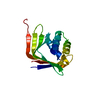
| ||||||||
|---|---|---|---|---|---|---|---|---|---|
| 1 | 
| ||||||||
| 単位格子 |
|
-要素
| #1: タンパク質 | 分子量: 22607.703 Da / 分子数: 1 / 断片: UNP residues 50-236 / 由来タイプ: 組換発現 / 由来: (組換発現) Mus musculus (ハツカネズミ) / 遺伝子: Ldb1, Nli / プラスミド: pHIS-GB1詳細 (発現宿主): T7 regulated expression with a His tag for purification and a GB1 tag to enhance soluble expression 発現宿主:  Escherichia coli BL21(DE3) (大腸菌) / 参照: UniProt: P70662 Escherichia coli BL21(DE3) (大腸菌) / 参照: UniProt: P70662 |
|---|---|
| #2: 水 | ChemComp-HOH / 水 分子量: 18.015 Da / 分子数: 9 / 由来タイプ: 天然 / 式: H2O 分子量: 18.015 Da / 分子数: 9 / 由来タイプ: 天然 / 式: H2O |
-実験情報
-実験
| 実験 | 手法: X線回折 / 使用した結晶の数: 2 |
|---|
- 試料調製
試料調製
| 結晶 |
| ||||||||||||
|---|---|---|---|---|---|---|---|---|---|---|---|---|---|
| 結晶化 |
|
-データ収集
| 回折 |
| ||||||||||||||||||||||||||||||||||||
|---|---|---|---|---|---|---|---|---|---|---|---|---|---|---|---|---|---|---|---|---|---|---|---|---|---|---|---|---|---|---|---|---|---|---|---|---|---|
| 放射光源 |
| ||||||||||||||||||||||||||||||||||||
| 検出器 |
| ||||||||||||||||||||||||||||||||||||
| 放射 |
| ||||||||||||||||||||||||||||||||||||
| 放射波長 |
| ||||||||||||||||||||||||||||||||||||
| 反射 | Entry-ID: 6PTL
| ||||||||||||||||||||||||||||||||||||
| 反射 シェル | Diffraction-ID: 1
|
- 解析
解析
| ソフトウェア |
| ||||||||||||||||||||||||||||||||||||||||||||||||||||||||||||
|---|---|---|---|---|---|---|---|---|---|---|---|---|---|---|---|---|---|---|---|---|---|---|---|---|---|---|---|---|---|---|---|---|---|---|---|---|---|---|---|---|---|---|---|---|---|---|---|---|---|---|---|---|---|---|---|---|---|---|---|---|---|
| 精密化 | 構造決定の手法: 分子置換 / 解像度: 2.5→35.48 Å / Cor.coef. Fo:Fc: 0.943 / Cor.coef. Fo:Fc free: 0.939 / SU B: 11.32 / SU ML: 0.234 / 交差検証法: THROUGHOUT / σ(F): 0 / ESU R: 0.357 / ESU R Free: 0.26 詳細: HYDROGENS HAVE BEEN ADDED IN THE RIDING POSITIONS U VALUES : REFINED INDIVIDUALLY
| ||||||||||||||||||||||||||||||||||||||||||||||||||||||||||||
| 溶媒の処理 | イオンプローブ半径: 0.8 Å / 減衰半径: 0.8 Å / VDWプローブ半径: 1.2 Å | ||||||||||||||||||||||||||||||||||||||||||||||||||||||||||||
| 原子変位パラメータ | Biso max: 163.85 Å2 / Biso mean: 87.291 Å2 / Biso min: 64.86 Å2
| ||||||||||||||||||||||||||||||||||||||||||||||||||||||||||||
| 精密化ステップ | サイクル: final / 解像度: 2.5→35.48 Å
| ||||||||||||||||||||||||||||||||||||||||||||||||||||||||||||
| 拘束条件 |
| ||||||||||||||||||||||||||||||||||||||||||||||||||||||||||||
| LS精密化 シェル | 解像度: 2.502→2.567 Å / Rfactor Rfree error: 0 / Total num. of bins used: 20
|
