 Movie
Movie Controller

[English] 日本語
 Yorodumi
- PDB-6ptl: Structure of the self-association domain of the chromatin looping... -
+ Open data

- Basic information
| Entry | Database: PDB / ID: 6ptl | |||||||||
|---|---|---|---|---|---|---|---|---|---|---|
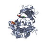
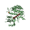
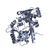
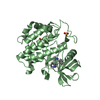
| Title | Structure of the self-association domain of the chromatin looping factor LDB1 | |||||||||
 Components Components | LIM domain-binding protein 1 | |||||||||
 Keywords Keywords |  GENE REGULATION / Dimer / Chromatin looping factor / nuclear protein / transcriptional regulator GENE REGULATION / Dimer / Chromatin looping factor / nuclear protein / transcriptional regulator | |||||||||
| Function / homology |  Function and homology informationregulation of kinase activity / cellular component assembly / negative regulation of erythrocyte differentiation / positive regulation of hemoglobin biosynthetic process / cerebellar Purkinje cell differentiation / epithelial structure maintenance / head development / primitive erythrocyte differentiation / transcription-dependent tethering of RNA polymerase II gene DNA at nuclear periphery / beta-catenin-TCF complex ...regulation of kinase activity / cellular component assembly / negative regulation of erythrocyte differentiation / positive regulation of hemoglobin biosynthetic process / cerebellar Purkinje cell differentiation / epithelial structure maintenance / head development / primitive erythrocyte differentiation / transcription-dependent tethering of RNA polymerase II gene DNA at nuclear periphery / beta-catenin-TCF complex / RUNX1 regulates transcription of genes involved in differentiation of HSCs / LIM domain binding / gastrulation with mouth forming second / anterior/posterior axis specification / regulation of focal adhesion assembly / cell leading edge / somatic stem cell population maintenance / hair follicle development / positive regulation of cell adhesion / regulation of cell migration / cerebellum development / positive regulation of transcription elongation by RNA polymerase II / neuron differentiation / Wnt signaling pathway / RNA polymerase II transcription regulator complex / : / nervous system development / DNA-binding transcription factor binding / transcription regulator complex / RNA polymerase II-specific DNA-binding transcription factor binding / transcription by RNA polymerase II / transcription coactivator activity / cell adhesion / negative regulation of DNA-templated transcription / chromatin binding / chromatin / negative regulation of transcription by RNA polymerase II / enzyme binding / protein homodimerization activity / positive regulation of transcription by RNA polymerase II / protein-containing complex / nucleoplasm / identical protein binding / nucleus Function and homology informationregulation of kinase activity / cellular component assembly / negative regulation of erythrocyte differentiation / positive regulation of hemoglobin biosynthetic process / cerebellar Purkinje cell differentiation / epithelial structure maintenance / head development / primitive erythrocyte differentiation / transcription-dependent tethering of RNA polymerase II gene DNA at nuclear periphery / beta-catenin-TCF complex ...regulation of kinase activity / cellular component assembly / negative regulation of erythrocyte differentiation / positive regulation of hemoglobin biosynthetic process / cerebellar Purkinje cell differentiation / epithelial structure maintenance / head development / primitive erythrocyte differentiation / transcription-dependent tethering of RNA polymerase II gene DNA at nuclear periphery / beta-catenin-TCF complex / RUNX1 regulates transcription of genes involved in differentiation of HSCs / LIM domain binding / gastrulation with mouth forming second / anterior/posterior axis specification / regulation of focal adhesion assembly / cell leading edge / somatic stem cell population maintenance / hair follicle development / positive regulation of cell adhesion / regulation of cell migration / cerebellum development / positive regulation of transcription elongation by RNA polymerase II / neuron differentiation / Wnt signaling pathway / RNA polymerase II transcription regulator complex / : / nervous system development / DNA-binding transcription factor binding / transcription regulator complex / RNA polymerase II-specific DNA-binding transcription factor binding / transcription by RNA polymerase II / transcription coactivator activity / cell adhesion / negative regulation of DNA-templated transcription / chromatin binding / chromatin / negative regulation of transcription by RNA polymerase II / enzyme binding / protein homodimerization activity / positive regulation of transcription by RNA polymerase II / protein-containing complex / nucleoplasm / identical protein binding / nucleusSimilarity search - Function | |||||||||
| Biological species |  Mus musculus (house mouse) Mus musculus (house mouse) | |||||||||
| Method | X-RAY DIFFRACTION / SYNCHROTRON / MOLECULAR REPLACEMENT / Resolution: 2.5 Å | |||||||||
 Authors Authors | Macindoe, I. / Silva, A. / Guss, J.M. / Mackay, J.P. / Matthews, J.M. | |||||||||
| Funding support |  Australia, 2items Australia, 2items
| |||||||||
 Citation Citation | Journal: To Be Published Title: Structure of the self-association domain of the chromatin looping factor LDB1 Authors: Macindoe, I. / Silva, A. / Guss, J.M. / Mackay, J.P. / Matthews, J.M. | |||||||||
| History |
|
- Structure visualization
| Structure viewer | Molecule: MolmilJmol/JSmol |
|---|
- Downloads & links
-Download
| PDBx/mmCIF format | 6ptl.cif.gz | 45.5 KB | Display | PDBx/mmCIF format |
|---|---|---|---|---|
| PDB format | pdb6ptl.ent.gz | 30.2 KB | Display | PDB format |
| PDBx/mmJSON format | 6ptl.json.gz | Tree view | PDBx/mmJSON format | |
| Others |  Other downloads Other downloads |
-Validation report
| Arichive directory | https://data.pdbj.org/pub/pdb/validation_reports/pt/6ptlftp://data.pdbj.org/pub/pdb/validation_reports/pt/6ptl | HTTPS FTP |
|---|
-Related structure data
| Similar structure data | |
|---|---|
| Other databases |
|


-Links
 PDBj
PDBj

- Assembly
| Deposited unit | 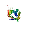
| ||||||||
|---|---|---|---|---|---|---|---|---|---|
| 1 | 
| ||||||||
| Unit cell |
|
-Components
| #1: Protein | Mass: 22607.703 Da / Num. of mol.: 1 / Fragment: UNP residues 50-236 Source method: isolated from a genetically manipulated source Source: (gene. exp.) Mus musculus (house mouse) / Gene: Ldb1, Nli / Plasmid: pHIS-GB1Details (production host): T7 regulated expression with a His tag for purification and a GB1 tag to enhance soluble expression Production host:  Escherichia coli BL21(DE3) (bacteria) / References: UniProt: P70662 Escherichia coli BL21(DE3) (bacteria) / References: UniProt: P70662 |
|---|---|
| #2: Water | ChemComp-HOH / Water Mass: 18.015 Da / Num. of mol.: 9 / Source method: isolated from a natural source / Formula: H2O Mass: 18.015 Da / Num. of mol.: 9 / Source method: isolated from a natural source / Formula: H2O |
-Experimental details
-Experiment
| Experiment | Method: X-RAY DIFFRACTION / Number of used crystals: 2 |
|---|
- Sample preparation
| Crystal |
| ||||||||||||
|---|---|---|---|---|---|---|---|---|---|---|---|---|---|
| Crystal grow |
|
-Data collection
| Diffraction |
| ||||||||||||||||||||||||||||||||||||
|---|---|---|---|---|---|---|---|---|---|---|---|---|---|---|---|---|---|---|---|---|---|---|---|---|---|---|---|---|---|---|---|---|---|---|---|---|---|
| Diffraction source |
| ||||||||||||||||||||||||||||||||||||
| Detector |
| ||||||||||||||||||||||||||||||||||||
| Radiation |
| ||||||||||||||||||||||||||||||||||||
| Radiation wavelength |
| ||||||||||||||||||||||||||||||||||||
| Reflection | Entry-ID: 6PTL
| ||||||||||||||||||||||||||||||||||||
| Reflection shell | Diffraction-ID: 1
|
- Processing
| Software |
| ||||||||||||||||||||||||||||||||||||||||||||||||||||||||||||
|---|---|---|---|---|---|---|---|---|---|---|---|---|---|---|---|---|---|---|---|---|---|---|---|---|---|---|---|---|---|---|---|---|---|---|---|---|---|---|---|---|---|---|---|---|---|---|---|---|---|---|---|---|---|---|---|---|---|---|---|---|---|
| Refinement | Method to determine structure: MOLECULAR REPLACEMENT / Resolution: 2.5→35.48 Å / Cor.coef. Fo:Fc: 0.943 / Cor.coef. Fo:Fc free: 0.939 / SU B: 11.32 / SU ML: 0.234 / Cross valid method: THROUGHOUT / σ(F): 0 / ESU R: 0.357 / ESU R Free: 0.26 Details: HYDROGENS HAVE BEEN ADDED IN THE RIDING POSITIONS U VALUES : REFINED INDIVIDUALLY
| ||||||||||||||||||||||||||||||||||||||||||||||||||||||||||||
| Solvent computation | Ion probe radii: 0.8 Å / Shrinkage radii: 0.8 Å / VDW probe radii: 1.2 Å | ||||||||||||||||||||||||||||||||||||||||||||||||||||||||||||
| Displacement parameters | Biso max: 163.85 Å2 / Biso mean: 87.291 Å2 / Biso min: 64.86 Å2
| ||||||||||||||||||||||||||||||||||||||||||||||||||||||||||||
| Refinement step | Cycle: final / Resolution: 2.5→35.48 Å
| ||||||||||||||||||||||||||||||||||||||||||||||||||||||||||||
| Refine LS restraints |
| ||||||||||||||||||||||||||||||||||||||||||||||||||||||||||||
| LS refinement shell | Resolution: 2.502→2.567 Å / Rfactor Rfree error: 0 / Total num. of bins used: 20
|
