Mass: 18.015 Da / Num. of mol.: 146 / Source method: isolated from a natural source / Formula: H2O
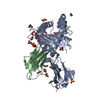
Has ligand of interest
N
-
Experimental details
-
Experiment
Experiment
Method: X-RAY DIFFRACTION / Number of used crystals: 1
-
Sample preparation
Crystal
Density Matthews: 2.26 Å3/Da / Density % sol: 45.68 %
Crystal grow
Temperature: 293 K / Method: batch mode / Details: 25-30% PEG6000, 50 mM imidazole / PH range: 7.5 - 8.0
-
Data collection
Diffraction
Mean temperature: 293 K / Ambient temp details: Room Temperature / Serial crystal experiment: N
Diffraction source
Source: SEALED TUBE / Type: OTHER / Wavelength: 1.5406 Å
Detector
Type: hilger and watts / Detector: DIFFRACTOMETER / Date: Feb 2, 1981
Radiation
Protocol: SINGLE WAVELENGTH / Monochromatic (M) / Laue (L): M / Scattering type: x-ray
Radiation wavelength
Wavelength: 1.5406 Å / Relative weight: 1
Reflection
Resolution: 2.26→39.05 Å / Num. obs: 16562 / % possible obs: 83.5 % / Redundancy: 5 % / Rmerge(I) obs: 0.068 / Net I/σ(I): 8.3
Reflection shell
Resolution: 2.26→2.87 Å / Num. unique obs: 6730 / % possible all: 67.8
-
Processing
Software
Name
Version
Classification
PHENIX
1.14_3247
refinement
PDB_EXTRACT
3.25
dataextraction
MOSFLM
datareduction
in-house
datascaling
in-house
phasing
Refinement
Method to determine structure: MIR / Resolution: 2.263→39.049 Å / SU ML: 0.2 / Cross valid method: THROUGHOUT / σ(F): 1.32 / Phase error: 19.49 / Stereochemistry target values: ML
Rfactor
Num. reflection
% reflection
Rfree
0.1961
783
4.73 %
Rwork
0.1538
15777
-
obs
0.1559
16560
83.51 %
Solvent computation
Shrinkage radii: 0.9 Å / VDW probe radii: 1.11 Å / Solvent model: FLAT BULK SOLVENT MODEL
In the structure databanks used in Yorodumi, some data are registered as the other names, "COVID-19 virus" and "2019-nCoV". Here are the details of the virus and the list of structure data.
Jan 31, 2019. EMDB accession codes are about to change! (news from PDBe EMDB page)
EMDB accession codes are about to change! (news from PDBe EMDB page)
The allocation of 4 digits for EMDB accession codes will soon come to an end. Whilst these codes will remain in use, new EMDB accession codes will include an additional digit and will expand incrementally as the available range of codes is exhausted. The current 4-digit format prefixed with “EMD-” (i.e. EMD-XXXX) will advance to a 5-digit format (i.e. EMD-XXXXX), and so on. It is currently estimated that the 4-digit codes will be depleted around Spring 2019, at which point the 5-digit format will come into force.
The EM Navigator/Yorodumi systems omit the EMD- prefix.
Related info.:Q: What is EMD? / ID/Accession-code notation in Yorodumi/EM Navigator
Yorodumi is a browser for structure data from EMDB, PDB, SASBDB, etc.
This page is also the successor to EM Navigator detail page, and also detail information page/front-end page for Omokage search.
The word "yorodu" (or yorozu) is an old Japanese word meaning "ten thousand". "mi" (miru) is to see.
Related info.:EMDB / PDB / SASBDB / Comparison of 3 databanks / Yorodumi Search / Aug 31, 2016. New EM Navigator & Yorodumi / Yorodumi Papers / Jmol/JSmol / Function and homology information / Changes in new EM Navigator and Yorodumi
 Movie
Movie Controller


 Open data
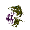
 Basic information
 Components
 Keywords
 Function and homology information

 Authors
 Citation
 Structure visualization
 Downloads & links
 Other downloads


 PDBj
PDBj
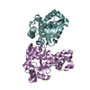
 Assembly

 Mass: 54.938 Da / Num. of mol.: 2 / Source method: obtained synthetically / Formula: Mn
 Mass: 18.015 Da / Num. of mol.: 146 / Source method: isolated from a natural source / Formula: H2O
 Sample preparation
 Processing