Movie
Movie Controller


+ Open data

- Basic information
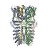
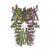
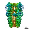
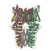
| Entry | Database: PDB / ID: 6pqq | ||||||
|---|---|---|---|---|---|---|---|
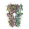
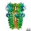
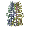
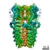
| Title | Cryo-EM structure of human TRPA1 C621S mutant in the apo state | ||||||
 Components Components | Transient receptor potential cation channel subfamily A member 1 | ||||||
 Keywords Keywords |  TRANSPORT PROTEIN / Ion channel / TRP channel / TRPA channel / TRPA1 channel / irritant sensing / apo state / membrane protein TRANSPORT PROTEIN / Ion channel / TRP channel / TRPA channel / TRPA1 channel / irritant sensing / apo state / membrane protein | ||||||
| Function / homology |  Function and homology information Function and homology informationtemperature-gated cation channel activity / stereocilium bundle / detection of chemical stimulus involved in sensory perception of pain / thermoception / TRP channels / channel activity / response to pain / cellular response to organic substance / intracellularly gated calcium channel activity / detection of mechanical stimulus involved in sensory perception of pain ...temperature-gated cation channel activity / stereocilium bundle / detection of chemical stimulus involved in sensory perception of pain / thermoception / TRP channels / channel activity / response to pain / cellular response to organic substance / intracellularly gated calcium channel activity / detection of mechanical stimulus involved in sensory perception of pain / monoatomic ion transport / sensory perception of pain / response to cold / response to organic substance / calcium ion transmembrane transport / calcium channel activity / intracellular calcium ion homeostasis / response to organic cyclic compound / cellular response to hydrogen peroxide / protein homotetramerization / cell surface receptor signaling pathway / response to xenobiotic stimulus / identical protein binding / plasma membraneSimilarity search - Function | ||||||
| Biological species |  Homo sapiens (human) Homo sapiens (human) | ||||||
| Method | ELECTRON MICROSCOPY / single particle reconstruction / cryo EM / Resolution: 2.81 Å | ||||||
 Authors Authors | Suo, Y. / Wang, Z. / Zubcevic, L. / Hsu, A.L. / He, Q. / Borgnia, M.J. / Ji, R.-R. / Lee, S.-Y. | ||||||
| Funding support |  United States, 1items United States, 1items
| ||||||
 Citation Citation | Journal: Neuron / Year: 2020 Title: Structural Insights into Electrophile Irritant Sensing by the Human TRPA1 Channel. Authors: Yang Suo / Zilong Wang / Lejla Zubcevic / Allen L Hsu / Qianru He / Mario J Borgnia / Ru-Rong Ji / Seok-Yong Lee / Abstract: Transient receptor potential channel subfamily A member 1 (TRPA1) is a Ca-permeable cation channel that serves as one of the primary sensors of environmental irritants and noxious substances. Many ...Transient receptor potential channel subfamily A member 1 (TRPA1) is a Ca-permeable cation channel that serves as one of the primary sensors of environmental irritants and noxious substances. Many TRPA1 agonists are electrophiles that are recognized by TRPA1 via covalent bond modifications of specific cysteine residues located in the cytoplasmic domains. However, a mechanistic understanding of electrophile sensing by TRPA1 has been limited due to a lack of high-resolution structural information. Here, we present the cryoelectron microscopy (cryo-EM) structures of nanodisc-reconstituted ligand-free TRPA1 and TRPA1 in complex with the covalent agonists JT010 and BITC at 2.8, 2.9, and 3.1 Å, respectively. Our structural and functional studies provide the molecular basis for electrophile recognition by the extraordinarily reactive C621 in TRPA1 and mechanistic insights into electrophile-dependent conformational changes in TRPA1. This work also provides a platform for future drug development targeting TRPA1. | ||||||
| History |
|
- Structure visualization
| Movie |
Movie viewer |
|---|---|
| Structure viewer | Molecule: MolmilJmol/JSmol |
- Downloads & links
-Download
| PDBx/mmCIF format | 6pqq.cif.gz | 491.9 KB | Display | PDBx/mmCIF format |
|---|---|---|---|---|
| PDB format | pdb6pqq.ent.gz | 376.7 KB | Display | PDB format |
| PDBx/mmJSON format | 6pqq.json.gz | Tree view | PDBx/mmJSON format | |
| Others |  Other downloads Other downloads |
-Validation report
| Arichive directory | https://data.pdbj.org/pub/pdb/validation_reports/pq/6pqqftp://data.pdbj.org/pub/pdb/validation_reports/pq/6pqq | HTTPS FTP |
|---|
-Related structure data
| Related structure data |  20451MC  6pqoC  6pqpC M: map data used to model this data C: citing same article ( |
|---|---|
| Similar structure data |


-Links
 PDBj
PDBj

- Assembly
| Deposited unit | 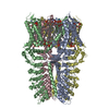
|
|---|---|
| 1 |
|
-Components
| #1: Protein | Mass: 131479.484 Da / Num. of mol.: 4 / Mutation: C621S Source method: isolated from a genetically manipulated source Source: (gene. exp.) Homo sapiens (human) / Gene: TRPA1, ANKTM1 / Production host: Homo sapiens (human) / References: UniProt: O75762#2: Chemical | ChemComp-LBN / POPC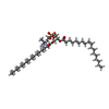  Mass: 760.076 Da / Num. of mol.: 8 / Source method: obtained synthetically / Formula: C42H82NO8P / Feature type: SUBJECT OF INVESTIGATION / Comment: phospholipid*YM Mass: 760.076 Da / Num. of mol.: 8 / Source method: obtained synthetically / Formula: C42H82NO8P / Feature type: SUBJECT OF INVESTIGATION / Comment: phospholipid*YM#3: Chemical | ChemComp-6OU / [( 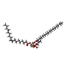  Mass: 717.996 Da / Num. of mol.: 20 / Source method: obtained synthetically / Formula: C39H76NO8P / Feature type: SUBJECT OF INVESTIGATION / Comment: phospholipid*YM Mass: 717.996 Da / Num. of mol.: 20 / Source method: obtained synthetically / Formula: C39H76NO8P / Feature type: SUBJECT OF INVESTIGATION / Comment: phospholipid*YMHas ligand of interest | Y | |
|---|
-Experimental details
-Experiment
| Experiment | Method: ELECTRON MICROSCOPY |
|---|---|
| EM experiment | Aggregation state: PARTICLE / 3D reconstruction method: single particle reconstruction |
- Sample preparation
| Component | Name: Transient receptor potential cation channel subfamily A member 1 Type: COMPLEX / Entity ID: #1 / Source: RECOMBINANT |
|---|---|
| Molecular weight | Experimental value: NO |
| Source (natural) | Organism: Homo sapiens (human) |
| Source (recombinant) | Organism: Homo sapiens (human) |
| Buffer solution | pH: 8 |
| Specimen | Conc.: 0.5 mg/ml / Embedding applied: NO / Shadowing applied: NO / Staining applied: NO / Vitrification applied: YES |
| Specimen support | Details: 15 mA / Grid material: GOLD / Grid mesh size: 300 divisions/in. / Grid type: UltrAuFoil |
| Vitrification | Instrument: LEICA EM GP / Cryogen name: ETHANE / Humidity: 100 % / Chamber temperature: 277 K |
- Electron microscopy imaging
| Experimental equipment |  Model: Titan Krios / Image courtesy: FEI Company |
|---|---|
| Microscopy | Model: FEI TITAN KRIOS |
| Electron gun | Electron source: FIELD EMISSION GUN / Accelerating voltage: 300 kV / Illumination mode: FLOOD BEAM |
| Electron lens | Mode: BRIGHT FIELDBright-field microscopy / Nominal magnification: 22500 X / Nominal defocus max: 2250 nm / Nominal defocus min: 750 nm / Cs: 2.7 mm / Alignment procedure: COMA FREE |
| Specimen holder | Cryogen: NITROGEN / Specimen holder model: FEI TITAN KRIOS AUTOGRID HOLDER |
| Image recording | Average exposure time: 4.6 sec. / Electron dose: 60 e/Å2 / Detector mode: COUNTING / Film or detector model: GATAN K3 (6k x 4k) / Num. of grids imaged: 1 / Num. of real images: 3544 |
- Processing
| Software | Name: PHENIX / Version: 1.13_2998: / Classification: refinement | ||||||||||||||||||||||||||||||||||||
|---|---|---|---|---|---|---|---|---|---|---|---|---|---|---|---|---|---|---|---|---|---|---|---|---|---|---|---|---|---|---|---|---|---|---|---|---|---|
| EM software |
| ||||||||||||||||||||||||||||||||||||
| CTF correction | Type: PHASE FLIPPING AND AMPLITUDE CORRECTION | ||||||||||||||||||||||||||||||||||||
| Particle selection | Num. of particles selected: 1150312 | ||||||||||||||||||||||||||||||||||||
| Symmetry | Point symmetry: C4 (4 fold cyclic) | ||||||||||||||||||||||||||||||||||||
| 3D reconstruction | Resolution: 2.81 Å / Resolution method: FSC 0.143 CUT-OFF / Num. of particles: 119697 / Num. of class averages: 1 / Symmetry type: POINT | ||||||||||||||||||||||||||||||||||||
| Atomic model building | B value: 100 / Protocol: RIGID BODY FIT / Space: REAL | ||||||||||||||||||||||||||||||||||||
| Atomic model building | PDB-ID: 3J9P Accession code: 3J9P / Source name: PDB / Type: experimental model | ||||||||||||||||||||||||||||||||||||
| Refinement | Highest resolution: 2.81 Å |