 Movie
Movie Controller


+ Open data

- Basic information
| Entry | Database: PDB / ID: 6ppo | ||||||
|---|---|---|---|---|---|---|---|
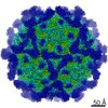
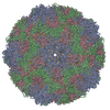
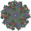
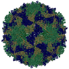
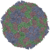
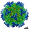
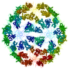
| Title | Rhinovirus C15 complexed with domain I of receptor CDHR3 | ||||||
 Components Components |
| ||||||
 Keywords Keywords | VIRUS/CELL ADHESION /  receptor / cadherin / VIRUS-CELL ADHESION complex receptor / cadherin / VIRUS-CELL ADHESION complex | ||||||
| Function / homology |  Function and homology information Function and homology informationcell-cell adhesion mediated by cadherin / calcium-dependent cell-cell adhesion via plasma membrane cell adhesion molecules / catenin complex / cell-cell junction assembly / adherens junction organization / homophilic cell adhesion via plasma membrane adhesion molecules / symbiont-mediated suppression of host cytoplasmic pattern recognition receptor signaling pathway via inhibition of RIG-I activity / synaptic cleft / axon terminus / picornain 2A ...cell-cell adhesion mediated by cadherin / calcium-dependent cell-cell adhesion via plasma membrane cell adhesion molecules / catenin complex / cell-cell junction assembly / adherens junction organization / homophilic cell adhesion via plasma membrane adhesion molecules / symbiont-mediated suppression of host cytoplasmic pattern recognition receptor signaling pathway via inhibition of RIG-I activity / synaptic cleft / axon terminus / picornain 2A / symbiont-mediated suppression of host mRNA export from nucleus / ribonucleoside triphosphate phosphatase activity / symbiont genome entry into host cell via pore formation in plasma membrane / picornain 3C / synaptic transmission, glutamatergic / T=pseudo3 icosahedral viral capsid / host cell cytoplasmic vesicle membrane / adherens junction / cell morphogenesis / endocytosis involved in viral entry into host cell / : / nucleoside-triphosphate phosphatase / virus receptor activity / protein complex oligomerization / monoatomic ion channel activity / RNA helicase activity / DNA replication / induction by virus of host autophagy / cadherin binding / RNA-directed RNA polymerase / symbiont-mediated suppression of host gene expression / viral RNA genome replication / cysteine-type endopeptidase activity / RNA-dependent RNA polymerase activity / DNA-templated transcription / host cell nucleus / calcium ion binding / virion attachment to host cell / structural molecule activity / proteolysis / RNA binding / ATP binding / membrane / metal ion binding / plasma membraneSimilarity search - Function | ||||||
| Biological species |  Homo sapiens (human) Homo sapiens (human) Rhinovirus C Rhinovirus C | ||||||
| Method | ELECTRON MICROSCOPY / single particle reconstruction / cryo EM / Resolution: 3.2 Å | ||||||
 Authors Authors | Sun, Y. / Watters, K. / Klose, T. / Palmenberg, A.C. | ||||||
| Funding support |  United States, 1items United States, 1items
| ||||||
 Citation Citation | Journal: Proc Natl Acad Sci U S A / Year: 2020 Title: Cryo-EM structure of rhinovirus C15a bound to its cadherin-related protein 3 receptor. Authors: Yingyuan Sun / Kelly Watters / Marchel G Hill / Qianglin Fang / Yue Liu / Richard J Kuhn / Thomas Klose / Michael G Rossmann / Ann C Palmenberg / Abstract: Infection by (RV-C), a species of Picornaviridae , is strongly associated with childhood asthma exacerbations. Cellular binding and entry by all RV-C, which trigger these episodes, is mediated by ...Infection by (RV-C), a species of Picornaviridae , is strongly associated with childhood asthma exacerbations. Cellular binding and entry by all RV-C, which trigger these episodes, is mediated by the first extracellular domain (EC1) of cadherin-related protein 3 (CDHR3), a surface cadherin-like protein expressed primarily on the apical surfaces of ciliated airway epithelial cells. Although recombinant EC1 is a potent inhibitor of viral infection, there is no molecular description of this protein or its binding site on RV-C. Here we present cryo-electron microscopy (EM) data resolving the EC1 and EC1+2 domains of human CDHR3 complexed with viral isolate C15a. Structure-suggested residues contributing to required interfaces on both EC1 and C15a were probed and identified by mutagenesis studies with four different RV-C genotypes. In contrast to most other rhinoviruses, which bind intercellular adhesion molecule 1 receptors via a capsid protein VP1-specific fivefold canyon feature, the CDHR3 EC1 contacts C15a, and presumably all RV-Cs, in a unique cohesive footprint near the threefold vertex, encompassing residues primarily from viral protein VP3, but also from VP1 and VP2. The EC1+2 footprint on C15a is similar to that of EC1 alone but shows that steric hindrance imposed by EC2 would likely prevent multiprotein binding by the native receptor at any singular threefold vertex. Definition of the molecular interface between the RV-Cs and their receptors provides new avenues that can be explored for potential antiviral therapies. | ||||||
| History |
|
- Structure visualization
| Movie |
Movie viewer |
|---|---|
| Structure viewer | Molecule: MolmilJmol/JSmol |
- Downloads & links
-Download
| PDBx/mmCIF format | 6ppo.cif.gz | 171 KB | Display | PDBx/mmCIF format |
|---|---|---|---|---|
| PDB format | pdb6ppo.ent.gz | 132.3 KB | Display | PDB format |
| PDBx/mmJSON format | 6ppo.json.gz | Tree view | PDBx/mmJSON format | |
| Others |  Other downloads Other downloads |
-Validation report
| Arichive directory | https://data.pdbj.org/pub/pdb/validation_reports/pp/6ppoftp://data.pdbj.org/pub/pdb/validation_reports/pp/6ppo | HTTPS FTP |
|---|
-Related structure data
| Related structure data | 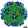 20443MC  6psfC M: map data used to model this data C: citing same article ( |
|---|---|
| Similar structure data |


-Links
 PDBj
PDBj


- Assembly
| Deposited unit | 
|
|---|---|
| 1 | x 60
|
| 2 |
|
| 3 | x 5
|
| 4 | x 6
|
| 5 | 
|
| Symmetry | Point symmetry: (Schoenflies symbol: I (icosahedral)) |
-Components
-Capsid protein ... , 4 types, 4 molecules ABCD
| #1: Protein | Mass: 31802.623 Da / Num. of mol.: 1 / Fragment: UNP residues 568-846 / Source method: isolated from a natural source / Source: (natural) Rhinovirus CReferences: UniProt: E5D8F2, picornain 2A, nucleoside-triphosphate phosphatase, picornain 3C, RNA-directed RNA polymerase |
|---|---|
| #2: Protein | Mass: 25965.037 Da / Num. of mol.: 1 / Fragment: UNP residues 333-567 / Source method: isolated from a natural source / Source: (natural) Rhinovirus CReferences: UniProt: E5D8F2, picornain 2A, nucleoside-triphosphate phosphatase, picornain 3C, RNA-directed RNA polymerase |
| #3: Protein | Mass: 29090.658 Da / Num. of mol.: 1 / Fragment: UNP residues 68-332 / Source method: isolated from a natural source / Source: (natural) Rhinovirus CReferences: UniProt: E5D8F2, picornain 2A, nucleoside-triphosphate phosphatase, picornain 3C, RNA-directed RNA polymerase |
| #4: Protein | Mass: 7174.758 Da / Num. of mol.: 1 / Fragment: UNP residues 2-67 / Source method: isolated from a natural source / Source: (natural) Rhinovirus CReferences: UniProt: E5D8F2, picornain 2A, nucleoside-triphosphate phosphatase, picornain 3C, RNA-directed RNA polymerase |
-Protein / Non-polymers , 2 types, 4 molecules U
| #5: Protein | Mass: 14517.157 Da / Num. of mol.: 1 Fragment: Extracellular cadherin-like domain 1 (UNP residues 20-130) Source method: isolated from a genetically manipulated source Source: (gene. exp.) Homo sapiens (human) / Gene: CDHR3, CDH28 / Production host:  Escherichia coli (E. coli) / References: UniProt: Q6ZTQ4 Escherichia coli (E. coli) / References: UniProt: Q6ZTQ4 |
|---|---|
| #6: Chemical |  Mass: 40.078 Da / Num. of mol.: 3 / Source method: obtained synthetically / Formula: Ca Mass: 40.078 Da / Num. of mol.: 3 / Source method: obtained synthetically / Formula: Ca |
-Details
| Has ligand of interest | N |
|---|
-Experimental details
-Experiment
| Experiment | Method: ELECTRON MICROSCOPY |
|---|---|
| EM experiment | Aggregation state: PARTICLE / 3D reconstruction method: single particle reconstruction |
- Sample preparation
| Component |
| ||||||||||||||||||||||||
|---|---|---|---|---|---|---|---|---|---|---|---|---|---|---|---|---|---|---|---|---|---|---|---|---|---|
| Molecular weight | Value: 6 MDa / Experimental value: NO | ||||||||||||||||||||||||
| Source (natural) |
| ||||||||||||||||||||||||
| Source (recombinant) | Organism: Escherichia coli (E. coli) | ||||||||||||||||||||||||
| Details of virus | Empty: NO / Enveloped: NO / Isolate: STRAIN / Type: VIRION | ||||||||||||||||||||||||
| Natural host | Organism: Homo sapiens | ||||||||||||||||||||||||
| Buffer solution | pH: 7.4 | ||||||||||||||||||||||||
| Specimen | Conc.: 0.5 mg/ml / Embedding applied: NO / Shadowing applied: NO / Staining applied: NO / Vitrification applied: YES | ||||||||||||||||||||||||
| Specimen support | Grid material: COPPER / Grid mesh size: 400 divisions/in. | ||||||||||||||||||||||||
| Vitrification | Instrument: GATAN CRYOPLUNGE 3 / Cryogen name: NITROGEN / Humidity: 80 % / Chamber temperature: 298 K / Details: 3s blotting time. Instrument placed in BSL2 hood. |
- Electron microscopy imaging
| Experimental equipment |  Model: Titan Krios / Image courtesy: FEI Company |
|---|---|
| Microscopy | Model: FEI TITAN KRIOS |
| Electron gun | Electron source: FIELD EMISSION GUN / Accelerating voltage: 300 kV / Illumination mode: FLOOD BEAM |
| Electron lens | Mode: BRIGHT FIELDBright-field microscopy / Nominal magnification: 81000 X / Nominal defocus max: 3000 nm / Nominal defocus min: 1000 nm / Cs: 2.7 mm / C2 aperture diameter: 100 µm / Alignment procedure: COMA FREE |
| Specimen holder | Cryogen: NITROGEN / Specimen holder model: FEI TITAN KRIOS AUTOGRID HOLDER |
| Image recording | Average exposure time: 12 sec. / Electron dose: 32 e/Å2 / Detector mode: SUPER-RESOLUTION / Film or detector model: GATAN K2 SUMMIT (4k x 4k) / Num. of grids imaged: 1 / Num. of real images: 1500 |
| EM imaging optics | Energyfilter name: GIF Quantum LS / Energyfilter slit width: 20 eV |
| Image scans | Sampling size: 0.865 µm / Width: 3838 / Height: 3710 / Movie frames/image: 60 / Used frames/image: 1-60 |
- Processing
| Software | Name: PHENIX / Version: 1.14_3260: / Classification: refinement | ||||||||||||||||||||||||||||||||||||||||
|---|---|---|---|---|---|---|---|---|---|---|---|---|---|---|---|---|---|---|---|---|---|---|---|---|---|---|---|---|---|---|---|---|---|---|---|---|---|---|---|---|---|
| EM software |
| ||||||||||||||||||||||||||||||||||||||||
| CTF correction | Type: PHASE FLIPPING AND AMPLITUDE CORRECTION | ||||||||||||||||||||||||||||||||||||||||
| Particle selection | Num. of particles selected: 22000 | ||||||||||||||||||||||||||||||||||||||||
| 3D reconstruction | Resolution: 3.2 Å / Resolution method: FSC 0.143 CUT-OFF / Num. of particles: 14978 / Algorithm: BACK PROJECTION / Symmetry type: POINT | ||||||||||||||||||||||||||||||||||||||||
| Atomic model building | B value: 100 / Protocol: AB INITIO MODEL / Space: REAL | ||||||||||||||||||||||||||||||||||||||||
| Atomic model building | PDB-ID: 5K0U Pdb chain-ID: A / Accession code: 5K0U / Source name: PDB / Type: experimental model |

