 Movie
Movie Controller

[English] 日本語
 Yorodumi
- PDB-6pom: Cryo-EM structure of the full-length Bacillus subtilis glyQS T-bo... -
+ Open data

- Basic information
| Entry | Database: PDB / ID: 6pom | ||||||
|---|---|---|---|---|---|---|---|
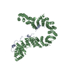
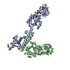
| Title | Cryo-EM structure of the full-length Bacillus subtilis glyQS T-box riboswitch in complex with tRNA-Gly | ||||||
 Components Components |
| ||||||
 Keywords Keywords |  RNA / RNA complex / riboswitch / transcription attenuation / stacking. RNA / RNA complex / riboswitch / transcription attenuation / stacking. | ||||||
| Function / homology | : / : / RNA / RNA (> 10) / RNA (> 100) Function and homology information Function and homology information | ||||||
| Biological species |  Bacillus subtilis (bacteria) Bacillus subtilis (bacteria) | ||||||
| Method | ELECTRON MICROSCOPY / single particle reconstruction / cryo EM / Resolution: 4.9 Å | ||||||
 Authors Authors | Li, S. / Su, Z. / Zhang, J. / Chiu, W. | ||||||
| Funding support |  United States, 1items United States, 1items
| ||||||
 Citation Citation | Journal: Nat Struct Mol Biol / Year: 2019 Title: Structural basis of amino acid surveillance by higher-order tRNA-mRNA interactions. Authors: Shuang Li / Zhaoming Su / Jean Lehmann / Vassiliki Stamatopoulou / Nikoleta Giarimoglou / Frances E Henderson / Lixin Fan / Grigore D Pintilie / Kaiming Zhang / Muyuan Chen / Steven J Ludtke ...Authors: Shuang Li / Zhaoming Su / Jean Lehmann / Vassiliki Stamatopoulou / Nikoleta Giarimoglou / Frances E Henderson / Lixin Fan / Grigore D Pintilie / Kaiming Zhang / Muyuan Chen / Steven J Ludtke / Yun-Xing Wang / Constantinos Stathopoulos / Wah Chiu / Jinwei Zhang /   Abstract: Amino acid availability in Gram-positive bacteria is monitored by T-box riboswitches. T-boxes directly bind tRNAs, assess their aminoacylation state, and regulate the transcription or translation of ...Amino acid availability in Gram-positive bacteria is monitored by T-box riboswitches. T-boxes directly bind tRNAs, assess their aminoacylation state, and regulate the transcription or translation of downstream genes to maintain nutritional homeostasis. Here, we report cocrystal and cryo-EM structures of Geobacillus kaustophilus and Bacillus subtilis T-box-tRNA complexes, detailing their multivalent, exquisitely selective interactions. The T-box forms a U-shaped molecular vise that clamps the tRNA, captures its 3' end using an elaborate 'discriminator' structure, and interrogates its aminoacylation state using a steric filter fashioned from a wobble base pair. In the absence of aminoacylation, T-boxes clutch tRNAs and form a continuously stacked central spine, permitting transcriptional readthrough or translation initiation. A modeled aminoacyl disrupts tRNA-T-box stacking, severing the central spine and blocking gene expression. Our data establish a universal mechanism of amino acid sensing on tRNAs and gene regulation by T-box riboswitches and exemplify how higher-order RNA-RNA interactions achieve multivalency and specificity. | ||||||
| History |
|
- Structure visualization
| Movie |
Movie viewer |
|---|---|
| Structure viewer | Molecule: MolmilJmol/JSmol |
- Downloads & links
-Download
| PDBx/mmCIF format | 6pom.cif.gz | 119.9 KB | Display | PDBx/mmCIF format |
|---|---|---|---|---|
| PDB format | pdb6pom.ent.gz | 86.3 KB | Display | PDB format |
| PDBx/mmJSON format | 6pom.json.gz | Tree view | PDBx/mmJSON format | |
| Others |  Other downloads Other downloads |
-Validation report
| Arichive directory | https://data.pdbj.org/pub/pdb/validation_reports/po/6pomftp://data.pdbj.org/pub/pdb/validation_reports/po/6pom | HTTPS FTP |
|---|
-Related structure data
| Related structure data | 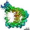 20416MC  6pmoC C: citing same article ( M: map data used to model this data |
|---|---|
| Similar structure data |


-Links
 PDBj
PDBj


- Assembly
| Deposited unit | 
|
|---|---|
| 1 |
|
-Components
| #1: RNA chain | Mass: 54730.551 Da / Num. of mol.: 1 Source method: isolated from a genetically manipulated source Source: (gene. exp.) Bacillus subtilis (bacteria) / Gene: glyQSProduction host: in vitro transcription vector pT7-TP(deltai) (others)References: GenBank: 2627063 |
|---|---|
| #2: RNA chain | Mass: 24156.336 Da / Num. of mol.: 1 Source method: isolated from a genetically manipulated source Source: (gene. exp.) Bacillus subtilis (bacteria)Production host: in vitro transcription vector pT7-TP(deltai) (others)References: GenBank: 1560688081 |
-Experimental details
-Experiment
| Experiment | Method: ELECTRON MICROSCOPY |
|---|---|
| EM experiment | Aggregation state: PARTICLE / 3D reconstruction method: single particle reconstruction |
- Sample preparation
| Component | Name: T-box-tRNAGly complex / Type: COMPLEX / Entity ID: all / Source: RECOMBINANT | ||||||||||||||||||||
|---|---|---|---|---|---|---|---|---|---|---|---|---|---|---|---|---|---|---|---|---|---|
| Molecular weight | Value: 0.075 MDa / Experimental value: NO | ||||||||||||||||||||
| Source (natural) | Organism: Bacillus subtilis (bacteria) | ||||||||||||||||||||
| Source (recombinant) | Organism: in vitro transcription vector pT7-TP(deltai) (others) | ||||||||||||||||||||
| Buffer solution | pH: 7.4 | ||||||||||||||||||||
| Buffer component |
| ||||||||||||||||||||
| Specimen | Conc.: 2.25 mg/ml / Embedding applied: NO / Shadowing applied: NO / Staining applied: NO / Vitrification applied: YES | ||||||||||||||||||||
| Specimen support | Grid material: COPPER / Grid mesh size: 200 divisions/in. / Grid type: Quantifoil R2/1 | ||||||||||||||||||||
| Vitrification | Instrument: FEI VITROBOT MARK IV / Cryogen name: ETHANE / Humidity: 100 % / Chamber temperature: 279 K / Details: 3 uL sample blot once for 3s |
- Electron microscopy imaging
| Experimental equipment |  Model: Titan Krios / Image courtesy: FEI Company |
|---|---|
| Microscopy | Model: FEI TITAN KRIOS |
| Electron gun | Electron source: FIELD EMISSION GUN / Accelerating voltage: 300 kV / Illumination mode: FLOOD BEAM |
| Electron lens | Mode: BRIGHT FIELDBright-field microscopy / Nominal magnification: 165000 X / Nominal defocus max: 3500 nm / Nominal defocus min: 400 nm / Cs: 2.7 mm / C2 aperture diameter: 70 µm / Alignment procedure: BASIC |
| Specimen holder | Cryogen: NITROGEN / Specimen holder model: FEI TITAN KRIOS AUTOGRID HOLDER / Temperature (max): 79 K / Temperature (min): 79 K |
| Image recording | Average exposure time: 5 sec. / Electron dose: 38 e/Å2 / Detector mode: COUNTING / Film or detector model: GATAN K2 QUANTUM (4k x 4k) / Num. of grids imaged: 3 / Num. of real images: 5600 |
| EM imaging optics | Energyfilter name: GIF Bioquantum / Energyfilter slit width: 20 eV |
| Image scans | Sampling size: 5 µm / Width: 3838 / Height: 3710 / Movie frames/image: 25 / Used frames/image: 1-25 |
- Processing
| Software | Name: PHENIX / Version: 1.15.2_3472: / Classification: refinement | ||||||||||||||||||||||||||||||||||||||||||||||||
|---|---|---|---|---|---|---|---|---|---|---|---|---|---|---|---|---|---|---|---|---|---|---|---|---|---|---|---|---|---|---|---|---|---|---|---|---|---|---|---|---|---|---|---|---|---|---|---|---|---|
| EM software |
| ||||||||||||||||||||||||||||||||||||||||||||||||
| Image processing | Details: Motion corrected by MotionCor2 | ||||||||||||||||||||||||||||||||||||||||||||||||
| CTF correction | Type: PHASE FLIPPING ONLY | ||||||||||||||||||||||||||||||||||||||||||||||||
| Particle selection | Num. of particles selected: 1962681 / Details: neural network particle picking module in EMAN2.2 | ||||||||||||||||||||||||||||||||||||||||||||||||
| Symmetry | Point symmetry: C1 (asymmetric) | ||||||||||||||||||||||||||||||||||||||||||||||||
| 3D reconstruction | Resolution: 4.9 Å / Resolution method: FSC 0.143 CUT-OFF / Num. of particles: 189361 / Algorithm: FOURIER SPACE / Num. of class averages: 2 / Symmetry type: POINT | ||||||||||||||||||||||||||||||||||||||||||||||||
| Atomic model building | Protocol: RIGID BODY FIT / Space: REAL / Target criteria: model map cross correlation | ||||||||||||||||||||||||||||||||||||||||||||||||
| Atomic model building | 3D fitting-ID: 1 / Accession code: 4LCK / Initial refinement model-ID: 1 / PDB-ID: 4LCK / Source name: PDB / Type: experimental model
| ||||||||||||||||||||||||||||||||||||||||||||||||
| Refine LS restraints |
|

