Movie
Movie Controller

[English] 日本語
 Yorodumi
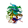
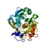
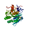
- PDB-6pks: MicroED structure of proteinase K from low-dose merged lamellae t... -
+ Open data

- Basic information
| Entry | Database: PDB / ID: 6pks | |||||||||
|---|---|---|---|---|---|---|---|---|---|---|
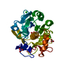
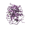
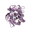
| Title | MicroED structure of proteinase K from low-dose merged lamellae that were not pre-coated with platinum 2.16A resolution (LD) | |||||||||
 Components Components | Proteinase K | |||||||||
 Keywords Keywords | HYDROLASE | |||||||||
| Function / homology |  Function and homology information Function and homology informationpeptidase K / serine-type endopeptidase activity / proteolysis / extracellular space / metal ion bindingSimilarity search - Function | |||||||||
| Biological species |  Parengyodontium album (fungus) Parengyodontium album (fungus) | |||||||||
| Method | ELECTRON CRYSTALLOGRAPHY / electron crystallography / cryo EM / Resolution: 2.16 Å | |||||||||
 Authors Authors | Martynowycz, M.W. / Zhao, W. / Hattne, J. / Jensen, G.J. / Gonen, T. | |||||||||
| Funding support |  United States, 2items United States, 2items
| |||||||||
 Citation Citation | Journal: Structure / Year: 2019 Title: Qualitative Analyses of Polishing and Precoating FIB Milled Crystals for MicroED. Authors: Michael W Martynowycz / Wei Zhao / Johan Hattne / Grant J Jensen / Tamir Gonen / Abstract: Microcrystal electron diffraction (MicroED) leverages the strong interaction between matter and electrons to determine protein structures from vanishingly small crystals. This strong interaction ...Microcrystal electron diffraction (MicroED) leverages the strong interaction between matter and electrons to determine protein structures from vanishingly small crystals. This strong interaction limits the thickness of crystals that can be investigated by MicroED, mainly due to absorption. Recent studies have demonstrated that focused ion-beam (FIB) milling can thin crystals into ideal-sized lamellae; however, it is not clear how to best apply FIB milling for MicroED. Here, the effects of polishing the lamellae, whereby the last few nanometers are milled away using a low-current gallium beam, are explored in both the platinum-precoated and uncoated samples. Our results suggest that precoating samples with a thin layer of platinum followed by polishing the crystal surfaces prior to data collection consistently led to superior results as indicated by higher signal-to-noise ratio, higher resolution, and better refinement statistics. This study lays the foundation for routine and reproducible methodology for sample preparation in MicroED. | |||||||||
| History |
|
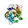
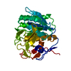
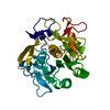
- Structure visualization
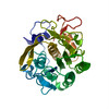
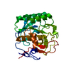
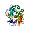
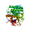
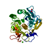
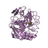
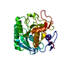
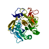
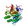
| Movie |
Movie viewer |
|---|---|
| Structure viewer | Molecule: MolmilJmol/JSmol |
- Downloads & links
-Download
| PDBx/mmCIF format | 6pks.cif.gz | 61.5 KB | Display | PDBx/mmCIF format |
|---|---|---|---|---|
| PDB format | pdb6pks.ent.gz | 45.9 KB | Display | PDB format |
| PDBx/mmJSON format | 6pks.json.gz | Tree view | PDBx/mmJSON format | |
| Others |  Other downloads Other downloads |
-Validation report
| Arichive directory | https://data.pdbj.org/pub/pdb/validation_reports/pk/6pksftp://data.pdbj.org/pub/pdb/validation_reports/pk/6pks | HTTPS FTP |
|---|
-Related structure data
| Related structure data |  20365MC 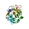 6pkjC 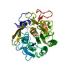 6pkkC 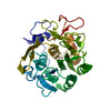 6pklC 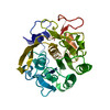 6pkmC 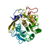 6pknC 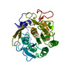 6pkoC 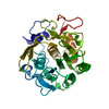 6pkpC 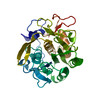 6pkqC  6pkrC 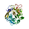 6pktC M: map data used to model this data C: citing same article ( |
|---|---|
| Similar structure data |


-Links
 PDBj
PDBj

- Assembly
| Deposited unit | 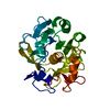
| ||||||||
|---|---|---|---|---|---|---|---|---|---|
| 1 |
| ||||||||
| Unit cell |
|
-Components
| #1: Protein | / Endopeptidase K / Tritirachium alkaline proteinase Mass: 28930.783 Da / Num. of mol.: 1 / Fragment: UNP residues 106-384 / Source method: isolated from a natural source / Source: (natural) Parengyodontium album (fungus) / References: UniProt: P06873, peptidase K |
|---|---|
| #2: Water | ChemComp-HOH / Water Mass: 18.015 Da / Num. of mol.: 55 / Source method: isolated from a natural source / Formula: H2O Mass: 18.015 Da / Num. of mol.: 55 / Source method: isolated from a natural source / Formula: H2O |
-Experimental details
-Experiment
| Experiment | Method: ELECTRON CRYSTALLOGRAPHY |
|---|---|
| EM experiment | Aggregation state: 3D ARRAY / 3D reconstruction method: electron crystallography |
- Sample preparation
| Component | Name: Proteinase K / Type: COMPLEX / Entity ID: #1 / Source: NATURAL |
|---|---|
| Molecular weight | Value: 0.02893 MDa / Experimental value: NO |
| Source (natural) | Organism: Parengyodontium album (fungus) |
| Buffer solution | pH: 7.5 |
| Specimen | Conc.: 20 mg/ml / Embedding applied: NO / Shadowing applied: NO / Staining applied: NO / Vitrification applied: YES / Details: Proteinase K purchased from Sigma. |
| Specimen support | Details: unspecified |
| Vitrification | Instrument: FEI VITROBOT MARK IV / Cryogen name: ETHANE / Humidity: 100 % / Chamber temperature: 273 K |
-Data collection
| Experimental equipment |  Model: Talos Arctica / Image courtesy: FEI Company |
|---|---|
| Microscopy | Model: FEI TALOS ARCTICA |
| Electron gun | Electron source: FIELD EMISSION GUN / Accelerating voltage: 200 kV / Illumination mode: FLOOD BEAM |
| Electron lens | Mode: DIFFRACTION / C2 aperture diameter: 100 µm / Alignment procedure: BASIC |
| Specimen holder | Cryogen: NITROGEN / Specimen holder model: FEI TITAN KRIOS AUTOGRID HOLDER / Temperature (max): 90 K / Temperature (min): 77 K |
| Image recording | Average exposure time: 3 sec. / Electron dose: 0.03 e/Å2 / Film or detector model: FEI CETA (4k x 4k) / Num. of diffraction images: 100 / Num. of grids imaged: 1 / Num. of real images: 100 Details: Continuous rotation with a rotation rate of 0.2 degrees per second and a readout every 3 seconds |
| Image scans | Sampling size: 28 µm / Width: 4096 / Height: 4096 |
| EM diffraction | Camera length: 2055 mm |
| EM diffraction shell | Resolution: 2.16→41.52 Å / Fourier space coverage: 98.87 % / Multiplicity: 9.4 / Num. of structure factors: 13527 / Phase residual: 26.59 ° |
| EM diffraction stats | Fourier space coverage: 98.87 % / High resolution: 2.16 Å / Num. of intensities measured: 126914 / Num. of structure factors: 13527 / Phase error: 26.59 ° / Phase residual: 26.59 ° / Phase error rejection criteria: 0 / Rmerge: 0.45 / Rsym: 0.47 |
- Processing
| Software | Name: PHENIX / Version: (1.14_3260: ???) / Classification: refinement | ||||||||||||||||||||||||||||||||||||||||||||||||||||||||||||||||||||||||||||||||||||
|---|---|---|---|---|---|---|---|---|---|---|---|---|---|---|---|---|---|---|---|---|---|---|---|---|---|---|---|---|---|---|---|---|---|---|---|---|---|---|---|---|---|---|---|---|---|---|---|---|---|---|---|---|---|---|---|---|---|---|---|---|---|---|---|---|---|---|---|---|---|---|---|---|---|---|---|---|---|---|---|---|---|---|---|---|---|
| EM software |
| ||||||||||||||||||||||||||||||||||||||||||||||||||||||||||||||||||||||||||||||||||||
| Image processing | Details: This was the new CetaD | ||||||||||||||||||||||||||||||||||||||||||||||||||||||||||||||||||||||||||||||||||||
| EM 3D crystal entity | ∠α: 90 ° / ∠β: 90 ° / ∠γ: 90 ° / A: 67.5 Å / B: 67.5 Å / C: 105.4 Å / Space group name: 96 / Space group num: 96 | ||||||||||||||||||||||||||||||||||||||||||||||||||||||||||||||||||||||||||||||||||||
| CTF correction | Type: NONE | ||||||||||||||||||||||||||||||||||||||||||||||||||||||||||||||||||||||||||||||||||||
| 3D reconstruction | Resolution: 2.16 Å / Resolution method: DIFFRACTION PATTERN/LAYERLINES / Symmetry type: 3D CRYSTAL | ||||||||||||||||||||||||||||||||||||||||||||||||||||||||||||||||||||||||||||||||||||
| Atomic model building | B value: 34.2 / Protocol: OTHER / Space: RECIPROCAL | ||||||||||||||||||||||||||||||||||||||||||||||||||||||||||||||||||||||||||||||||||||
| Atomic model building | PDB-ID: 6CL7 Pdb chain-ID: A / Pdb chain residue range: 106-384 | ||||||||||||||||||||||||||||||||||||||||||||||||||||||||||||||||||||||||||||||||||||
| Refinement | Resolution: 2.16→41.52 Å / SU ML: 0.31 / Cross valid method: THROUGHOUT / σ(F): 1.34 / Phase error: 26.58
| ||||||||||||||||||||||||||||||||||||||||||||||||||||||||||||||||||||||||||||||||||||
| Solvent computation | Shrinkage radii: 0.9 Å / VDW probe radii: 1.11 Å | ||||||||||||||||||||||||||||||||||||||||||||||||||||||||||||||||||||||||||||||||||||
| Refine LS restraints |
| ||||||||||||||||||||||||||||||||||||||||||||||||||||||||||||||||||||||||||||||||||||
| LS refinement shell |
|