 Movie
Movie Controller

[English] 日本語
 Yorodumi
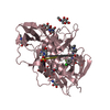
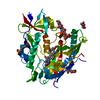
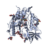
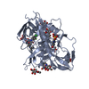
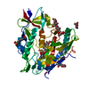
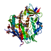
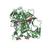
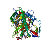
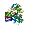
- PDB-6onv: Crystal structure of HIV-1 LM/HT Clade A/E CRF01 gp120 core in co... -
+ Open data

- Basic information
| Entry | Database: PDB / ID: 6onv | ||||||||||||
|---|---|---|---|---|---|---|---|---|---|---|---|---|---|
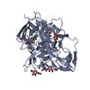
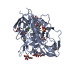
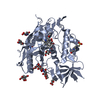
| Title | Crystal structure of HIV-1 LM/HT Clade A/E CRF01 gp120 core in complex with (S)-MCG-III-027-D05. | ||||||||||||
 Components Components | HIV-1 LM/HT Clade A/E CRF01 gp120 | ||||||||||||
 Keywords Keywords |  viral protein/inhibitor / HIV-1 gp120 / CLADE A/E CF01 / viral protein / viral protein-inhibitor complex viral protein/inhibitor / HIV-1 gp120 / CLADE A/E CF01 / viral protein / viral protein-inhibitor complex | ||||||||||||
| Function / homology | Gp120 core superfamily / Envelope glycoprotein GP120 / Human immunodeficiency virus 1, envelope glycoprotein Gp120 / viral envelope / Chem-MWA / clade A/E 93TH057 HIV-1 gp120 core Function and homology information Function and homology information | ||||||||||||
| Biological species |   Human immunodeficiency virus 1 Human immunodeficiency virus 1 | ||||||||||||
| Method | X-RAY DIFFRACTION / SYNCHROTRON / MOLECULAR REPLACEMENT / Resolution: 3.253 Å | ||||||||||||
 Authors Authors | Tolbert, W.D. / Sherburn, R. / Pazgier, M. | ||||||||||||
| Funding support |  United States, 3items United States, 3items
| ||||||||||||
 Citation Citation | Journal: J.Virol. / Year: 2019 Title: A New Family of Small-Molecule CD4-Mimetic Compounds Contacts Highly Conserved Aspartic Acid 368 of HIV-1 gp120 and Mediates Antibody-Dependent Cellular Cytotoxicity. Authors: Ding, S. / Grenier, M.C. / Tolbert, W.D. / Vezina, D. / Sherburn, R. / Richard, J. / Prevost, J. / Chapleau, J.P. / Gendron-Lepage, G. / Medjahed, H. / Abrams, C. / Sodroski, J. / Pazgier, M. ...Authors: Ding, S. / Grenier, M.C. / Tolbert, W.D. / Vezina, D. / Sherburn, R. / Richard, J. / Prevost, J. / Chapleau, J.P. / Gendron-Lepage, G. / Medjahed, H. / Abrams, C. / Sodroski, J. / Pazgier, M. / Smith 3rd, A.B. / Finzi, A. | ||||||||||||
| History |
|
- Structure visualization
| Structure viewer | Molecule: MolmilJmol/JSmol |
|---|
- Downloads & links
-Download
| PDBx/mmCIF format | 6onv.cif.gz | 88.2 KB | Display | PDBx/mmCIF format |
|---|---|---|---|---|
| PDB format | pdb6onv.ent.gz | 63.7 KB | Display | PDB format |
| PDBx/mmJSON format | 6onv.json.gz | Tree view | PDBx/mmJSON format | |
| Others |  Other downloads Other downloads |
-Validation report
| Arichive directory | https://data.pdbj.org/pub/pdb/validation_reports/on/6onvftp://data.pdbj.org/pub/pdb/validation_reports/on/6onv | HTTPS FTP |
|---|
-Related structure data
| Related structure data | 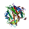 6oneC  6onfC 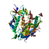 6onhC 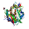 6p9nC  3tgtS S: Starting model for refinement C: citing same article ( |
|---|---|
| Similar structure data |


-Links
 PDBj
PDBj


- Assembly
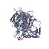
| Deposited unit | 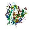
| ||||||||
|---|---|---|---|---|---|---|---|---|---|
| 1 |
| ||||||||
| Unit cell |
|
-Components
| #1: Protein | Mass: 39466.750 Da / Num. of mol.: 1 / Mutation: H61Y, Q105H, V108I, H375T, N474D, I475M, K476R Source method: isolated from a genetically manipulated source Source: (gene. exp.) Human immunodeficiency virus 1 / Gene: HIV-1 Env / Cell (production host): HEK GnT1- / Production host:  Homo sapiens (human) / References: UniProt: A0A0M3KKW9 Homo sapiens (human) / References: UniProt: A0A0M3KKW9 | ||||||
|---|---|---|---|---|---|---|---|
| #2: Sugar | ChemComp-NAG / N-Acetylglucosamine  Type: D-saccharide, beta linking / Mass: 221.208 Da / Num. of mol.: 10 Type: D-saccharide, beta linking / Mass: 221.208 Da / Num. of mol.: 10Source method: isolated from a genetically manipulated source Formula: C8H15NO6 #3: Chemical | ChemComp-MWA / ( |   Mass: 334.794 Da / Num. of mol.: 1 / Source method: obtained synthetically / Formula: C13H16ClFN2O3S / Feature type: SUBJECT OF INVESTIGATION Mass: 334.794 Da / Num. of mol.: 1 / Source method: obtained synthetically / Formula: C13H16ClFN2O3S / Feature type: SUBJECT OF INVESTIGATION#4: Chemical | ChemComp-EPE / | HEPES  Mass: 238.305 Da / Num. of mol.: 1 / Source method: obtained synthetically / Formula: C8H18N2O4S / Comment: pH buffer*YM Mass: 238.305 Da / Num. of mol.: 1 / Source method: obtained synthetically / Formula: C8H18N2O4S / Comment: pH buffer*YMHas ligand of interest | Y | |
-Experimental details
-Experiment
| Experiment | Method: X-RAY DIFFRACTION / Number of used crystals: 1 |
|---|
- Sample preparation
| Crystal | Density Matthews: 2.44 Å3/Da / Density % sol: 49.56 % |
|---|---|
| Crystal grow | Temperature: 294 K / Method: vapor diffusion, hanging drop / pH: 7.5 / Details: 10% PEG 1500 5% PEG 400 0.1M HEPES pH 7.5 |
-Data collection
| Diffraction | Mean temperature: 100 K / Serial crystal experiment: N |
|---|---|
| Diffraction source | Source: SYNCHROTRON / Site: SSRL / Beamline: BL12-2 / Wavelength: 0.97946 Å |
| Detector | Type: DECTRIS PILATUS 6M / Detector: PIXEL / Date: Nov 9, 2018 |
| Radiation | Monochromator: Si(111) / Protocol: SINGLE WAVELENGTH / Monochromatic (M) / Laue (L): M / Scattering type: x-ray |
| Radiation wavelength | Wavelength: 0.97946 Å / Relative weight: 1 |
| Reflection | Resolution: 3.25→50 Å / Num. obs: 4355 / % possible obs: 67.6 % / Redundancy: 4.4 % / CC1/2: 0.96 / Rmerge(I) obs: 0.213 / Rpim(I) all: 0.108 / Net I/σ(I): 12.6 |
| Reflection shell | Resolution: 3.25→3.31 Å / Redundancy: 4.1 % / Rmerge(I) obs: 0.866 / Mean I/σ(I) obs: 1.6 / Num. unique obs: 211 / CC1/2: 0.45 / Rpim(I) all: 0.513 / % possible all: 65.9 |
- Processing
| Software |
| ||||||||||||||||||||||||
|---|---|---|---|---|---|---|---|---|---|---|---|---|---|---|---|---|---|---|---|---|---|---|---|---|---|
| Refinement | Method to determine structure: MOLECULAR REPLACEMENT Starting model: 3TGT Resolution: 3.253→33.128 Å / SU ML: 0.29 / Cross valid method: FREE R-VALUE / σ(F): 1.38 / Phase error: 18.07
| ||||||||||||||||||||||||
| Solvent computation | Shrinkage radii: 0.9 Å / VDW probe radii: 1.11 Å | ||||||||||||||||||||||||
| Refinement step | Cycle: LAST / Resolution: 3.253→33.128 Å
| ||||||||||||||||||||||||
| Refine LS restraints |
| ||||||||||||||||||||||||
| LS refinement shell |
|
