 Movie
Movie Controller

[English] 日本語
 Yorodumi
- PDB-6ogn: Crystal structure of mouse protein arginine methyltransferase 7 i... -
+ Open data

- Basic information
| Entry | Database: PDB / ID: 6ogn | |||||||||
|---|---|---|---|---|---|---|---|---|---|---|
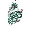
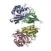
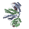
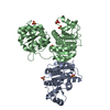
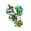
| Title | Crystal structure of mouse protein arginine methyltransferase 7 in complex with SGC8158 chemical probe | |||||||||
 Components Components | Protein arginine N-methyltransferase 7 | |||||||||
 Keywords Keywords |  TRANSFERASE / PRMT7 / SGC8158 / chemical probe / Structural Genomics / Structural Genomics Consortium TRANSFERASE / PRMT7 / SGC8158 / chemical probe / Structural Genomics / Structural Genomics Consortium | |||||||||
| Function / homology |  Function and homology information Function and homology informationtype III protein arginine methyltransferase / protein-arginine omega-N monomethyltransferase activity / histone H4 methyltransferase activity / protein-arginine omega-N symmetric methyltransferase activity / peptidyl-arginine methylation / histone H4R3 methyltransferase activity / RMTs methylate histone arginines / genomic imprinting / S-adenosylmethionine-dependent methyltransferase activity / spliceosomal snRNP assembly ...type III protein arginine methyltransferase / protein-arginine omega-N monomethyltransferase activity / histone H4 methyltransferase activity / protein-arginine omega-N symmetric methyltransferase activity / peptidyl-arginine methylation / histone H4R3 methyltransferase activity / RMTs methylate histone arginines / genomic imprinting / S-adenosylmethionine-dependent methyltransferase activity / spliceosomal snRNP assembly / ribonucleoprotein complex binding / fibrillar center / histone binding / nucleoplasm / nucleus / cytosolSimilarity search - Function | |||||||||
| Biological species |  Mus musculus (house mouse) Mus musculus (house mouse) | |||||||||
| Method | X-RAY DIFFRACTION / SYNCHROTRON / FOURIER SYNTHESIS / Resolution: 2.4 Å | |||||||||
 Authors Authors | Halabelian, L. / Dong, A. / Zeng, H. / Li, Y. / Hutchinson, A. / Seitova, A. / Bountra, C. / Edwards, A.M. / Arrowsmith, C.H. / Structural Genomics Consortium (SGC) | |||||||||
 Citation Citation | Journal: Nat Commun / Year: 2020 Title: Pharmacological inhibition of PRMT7 links arginine monomethylation to the cellular stress response. Authors: Szewczyk, M.M. / Ishikawa, Y. / Organ, S. / Sakai, N. / Li, F. / Halabelian, L. / Ackloo, S. / Couzens, A.L. / Eram, M. / Dilworth, D. / Fukushi, H. / Harding, R. / Dela Sena, C.C. / Sugo, T. ...Authors: Szewczyk, M.M. / Ishikawa, Y. / Organ, S. / Sakai, N. / Li, F. / Halabelian, L. / Ackloo, S. / Couzens, A.L. / Eram, M. / Dilworth, D. / Fukushi, H. / Harding, R. / Dela Sena, C.C. / Sugo, T. / Hayashi, K. / McLeod, D. / Zepeda, C. / Aman, A. / Sanchez-Osuna, M. / Bonneil, E. / Takagi, S. / Al-Awar, R. / Tyers, M. / Richard, S. / Takizawa, M. / Gingras, A.C. / Arrowsmith, C.H. / Vedadi, M. / Brown, P.J. / Nara, H. / Barsyte-Lovejoy, D. | |||||||||
| History |
|
- Structure visualization
| Structure viewer | Molecule: MolmilJmol/JSmol |
|---|
- Downloads & links
-Download
| PDBx/mmCIF format | 6ogn.cif.gz | 138.8 KB | Display | PDBx/mmCIF format |
|---|---|---|---|---|
| PDB format | pdb6ogn.ent.gz | 103.8 KB | Display | PDB format |
| PDBx/mmJSON format | 6ogn.json.gz | Tree view | PDBx/mmJSON format | |
| Others |  Other downloads Other downloads |
-Validation report
| Arichive directory | https://data.pdbj.org/pub/pdb/validation_reports/og/6ognftp://data.pdbj.org/pub/pdb/validation_reports/og/6ogn | HTTPS FTP |
|---|
-Related structure data
| Related structure data |  4c4aS S: Starting model for refinement |
|---|---|
| Similar structure data |

-Links
 PDBj
PDBj- Assembly
| Deposited unit | 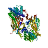
| ||||||||
|---|---|---|---|---|---|---|---|---|---|
| 1 |
| ||||||||
| Unit cell |
|
-Components
| #1: Protein | Mass: 78391.086 Da / Num. of mol.: 1 Source method: isolated from a genetically manipulated source Source: (gene. exp.) Mus musculus (house mouse) / Gene: Prmt7, Kiaa1933 / Plasmid: pFBOH-MHL / Production host:   Spodoptera frugiperda (fall armyworm) Spodoptera frugiperda (fall armyworm)References: UniProt: Q922X9, type III protein arginine methyltransferase | ||
|---|---|---|---|
| #2: Chemical | ChemComp-ZN /   Mass: 65.409 Da / Num. of mol.: 1 / Source method: obtained synthetically / Formula: Zn Mass: 65.409 Da / Num. of mol.: 1 / Source method: obtained synthetically / Formula: Zn | ||
| #3: Chemical | ChemComp-MJ7 /   Mass: 555.091 Da / Num. of mol.: 1 / Source method: obtained synthetically / Formula: C27H31ClN6O3S Mass: 555.091 Da / Num. of mol.: 1 / Source method: obtained synthetically / Formula: C27H31ClN6O3S | ||
| #4: Chemical |   Mass: 647.004 Da / Num. of mol.: 3 / Source method: obtained synthetically Mass: 647.004 Da / Num. of mol.: 3 / Source method: obtained synthetically#5: Water | ChemComp-HOH / | Water Mass: 18.015 Da / Num. of mol.: 42 / Source method: isolated from a natural source / Formula: H2O Mass: 18.015 Da / Num. of mol.: 42 / Source method: isolated from a natural source / Formula: H2O |
-Experimental details
-Experiment
| Experiment | Method: X-RAY DIFFRACTION / Number of used crystals: 1 |
|---|
- Sample preparation
| Crystal | Density Matthews: 2.59 Å3/Da / Density % sol: 52.44 % / Mosaicity: 0.418 ° |
|---|---|
| Crystal grow | Temperature: 293 K / Method: vapor diffusion, sitting drop / pH: 6.4 Details: 20% PEG3350, 0.04 M citric acid, 0.06 M Bis-Tris propane, pH 6.4 |
-Data collection
| Diffraction | Mean temperature: 100 K / Serial crystal experiment: N | |||||||||||||||||||||||||||||||||||||||||||||||||||||||||||||||||||||||||||||||||||||||||||||||||||||||||||||||||||||||||||||||||||||||||||||||||||||||||||||||||||||||||||||||||||||||||||||
|---|---|---|---|---|---|---|---|---|---|---|---|---|---|---|---|---|---|---|---|---|---|---|---|---|---|---|---|---|---|---|---|---|---|---|---|---|---|---|---|---|---|---|---|---|---|---|---|---|---|---|---|---|---|---|---|---|---|---|---|---|---|---|---|---|---|---|---|---|---|---|---|---|---|---|---|---|---|---|---|---|---|---|---|---|---|---|---|---|---|---|---|---|---|---|---|---|---|---|---|---|---|---|---|---|---|---|---|---|---|---|---|---|---|---|---|---|---|---|---|---|---|---|---|---|---|---|---|---|---|---|---|---|---|---|---|---|---|---|---|---|---|---|---|---|---|---|---|---|---|---|---|---|---|---|---|---|---|---|---|---|---|---|---|---|---|---|---|---|---|---|---|---|---|---|---|---|---|---|---|---|---|---|---|---|---|---|---|---|---|---|
| Diffraction source | Source: SYNCHROTRON / Site: APS  / Beamline: 24-ID-E / Wavelength: 0.97918 Å / Beamline: 24-ID-E / Wavelength: 0.97918 Å | |||||||||||||||||||||||||||||||||||||||||||||||||||||||||||||||||||||||||||||||||||||||||||||||||||||||||||||||||||||||||||||||||||||||||||||||||||||||||||||||||||||||||||||||||||||||||||||
| Detector | Type: DECTRIS EIGER X 16M / Detector: PIXEL / Date: Feb 20, 2019 | |||||||||||||||||||||||||||||||||||||||||||||||||||||||||||||||||||||||||||||||||||||||||||||||||||||||||||||||||||||||||||||||||||||||||||||||||||||||||||||||||||||||||||||||||||||||||||||
| Radiation | Monochromator: Cryogenically-cooled single crystal Si(220) side bounce Protocol: SINGLE WAVELENGTH / Monochromatic (M) / Laue (L): M / Scattering type: x-ray | |||||||||||||||||||||||||||||||||||||||||||||||||||||||||||||||||||||||||||||||||||||||||||||||||||||||||||||||||||||||||||||||||||||||||||||||||||||||||||||||||||||||||||||||||||||||||||||
| Radiation wavelength | Wavelength: 0.97918 Å / Relative weight: 1 | |||||||||||||||||||||||||||||||||||||||||||||||||||||||||||||||||||||||||||||||||||||||||||||||||||||||||||||||||||||||||||||||||||||||||||||||||||||||||||||||||||||||||||||||||||||||||||||
| Reflection | Resolution: 2.4→50 Å / Num. obs: 33363 / % possible obs: 99.7 % / Redundancy: 12.9 % / Rmerge(I) obs: 0.114 / Rpim(I) all: 0.033 / Rrim(I) all: 0.118 / Χ2: 1.919 / Net I/σ(I): 9.5 | |||||||||||||||||||||||||||||||||||||||||||||||||||||||||||||||||||||||||||||||||||||||||||||||||||||||||||||||||||||||||||||||||||||||||||||||||||||||||||||||||||||||||||||||||||||||||||||
| Reflection shell | Diffraction-ID: 1
|
- Processing
| Software |
| ||||||||||||||||||||||||||||||||||||||||||||||||||||||||||||||||||||||||||||||||||||||||||||||||||||||||||||||||||||||||||||||||||||||||||||||||||||||||||||||||||||||||||||||||||||||
|---|---|---|---|---|---|---|---|---|---|---|---|---|---|---|---|---|---|---|---|---|---|---|---|---|---|---|---|---|---|---|---|---|---|---|---|---|---|---|---|---|---|---|---|---|---|---|---|---|---|---|---|---|---|---|---|---|---|---|---|---|---|---|---|---|---|---|---|---|---|---|---|---|---|---|---|---|---|---|---|---|---|---|---|---|---|---|---|---|---|---|---|---|---|---|---|---|---|---|---|---|---|---|---|---|---|---|---|---|---|---|---|---|---|---|---|---|---|---|---|---|---|---|---|---|---|---|---|---|---|---|---|---|---|---|---|---|---|---|---|---|---|---|---|---|---|---|---|---|---|---|---|---|---|---|---|---|---|---|---|---|---|---|---|---|---|---|---|---|---|---|---|---|---|---|---|---|---|---|---|---|---|---|---|
| Refinement | Method to determine structure: FOURIER SYNTHESIS Starting model: PDB entry 4C4A Resolution: 2.4→47.03 Å / Cor.coef. Fo:Fc: 0.95 / Cor.coef. Fo:Fc free: 0.927 / SU B: 8.213 / SU ML: 0.192 / Cross valid method: THROUGHOUT / ESU R: 0.321 / ESU R Free: 0.25 / Stereochemistry target values: MAXIMUM LIKELIHOOD / Details: HYDROGENS HAVE BEEN ADDED IN THE RIDING POSITIONS
| ||||||||||||||||||||||||||||||||||||||||||||||||||||||||||||||||||||||||||||||||||||||||||||||||||||||||||||||||||||||||||||||||||||||||||||||||||||||||||||||||||||||||||||||||||||||
| Solvent computation | Ion probe radii: 0.8 Å / Shrinkage radii: 0.8 Å / VDW probe radii: 1.2 Å / Solvent model: MASK | ||||||||||||||||||||||||||||||||||||||||||||||||||||||||||||||||||||||||||||||||||||||||||||||||||||||||||||||||||||||||||||||||||||||||||||||||||||||||||||||||||||||||||||||||||||||
| Displacement parameters | Biso mean: 63.365 Å2
| ||||||||||||||||||||||||||||||||||||||||||||||||||||||||||||||||||||||||||||||||||||||||||||||||||||||||||||||||||||||||||||||||||||||||||||||||||||||||||||||||||||||||||||||||||||||
| Refinement step | Cycle: 1 / Resolution: 2.4→47.03 Å
| ||||||||||||||||||||||||||||||||||||||||||||||||||||||||||||||||||||||||||||||||||||||||||||||||||||||||||||||||||||||||||||||||||||||||||||||||||||||||||||||||||||||||||||||||||||||
| Refine LS restraints |
|