 Movie
Movie Controller

[English] 日本語
 Yorodumi
- PDB-6o9f: The structure of Thermomyces Lanuginosa lipase in complex with 1,... -
+ Open data

- Basic information
| Entry | Database: PDB / ID: 6o9f | ||||||
|---|---|---|---|---|---|---|---|
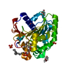
| Title | The structure of Thermomyces Lanuginosa lipase in complex with 1,3 diacylglycerol in a monoclinic crystal form | ||||||
 Components Components | Lipase | ||||||
 Keywords Keywords | HYDROLASE / lipids / triglycerides / catalytic triad / monolayer / acyl intermediate / trimer / NCS / LIPID BINDING PROTEIN | ||||||
| Function / homology |  Function and homology informationtriacylglycerol lipase / triglyceride lipase activity / lipid catabolic process Function and homology informationtriacylglycerol lipase / triglyceride lipase activity / lipid catabolic processSimilarity search - Function | ||||||
| Biological species |   Thermomyces lanuginosus (fungus) Thermomyces lanuginosus (fungus) | ||||||
| Method | X-RAY DIFFRACTION / MOLECULAR REPLACEMENT / Resolution: 2.48 Å | ||||||
 Authors Authors | McPherson, A. | ||||||
| Funding support | 1items
| ||||||
 Citation Citation | Journal: Curr Enzym Inhib / Year: 2020 Title: The Crystal Structures of Thermomyces (Humicola) Lanuginosa Lipase in Complex with Enzymatic Reactants Authors: McPherson , A. / Larson , B.S. / Kalasky , A. | ||||||
| History |
|
- Structure visualization
| Structure viewer | Molecule: MolmilJmol/JSmol |
|---|
- Downloads & links
-Download
| PDBx/mmCIF format | 6o9f.cif.gz | 738.4 KB | Display | PDBx/mmCIF format |
|---|---|---|---|---|
| PDB format | pdb6o9f.ent.gz | 500.9 KB | Display | PDB format |
| PDBx/mmJSON format | 6o9f.json.gz | Tree view | PDBx/mmJSON format | |
| Others |  Other downloads Other downloads |
-Validation report
| Arichive directory | https://data.pdbj.org/pub/pdb/validation_reports/o9/6o9fftp://data.pdbj.org/pub/pdb/validation_reports/o9/6o9f | HTTPS FTP |
|---|
-Related structure data
-Links
 PDBj
PDBj- Assembly
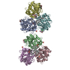
| Deposited unit | 
| ||||||||||
|---|---|---|---|---|---|---|---|---|---|---|---|
| 1 |
| ||||||||||
| 2 |
| ||||||||||
| Unit cell |
|
-Components
-Protein / Sugars , 2 types, 12 molecules ABCEDF
| #1: Protein | / Triacylglycerol lipase Mass: 31836.459 Da / Num. of mol.: 6 Source method: isolated from a genetically manipulated source Source: (gene. exp.) Thermomyces lanuginosus (fungus) / Gene: LIP / Production host:  Thermoactinomyces vulgaris (bacteria) / References: UniProt: O59952, triacylglycerol lipase Thermoactinomyces vulgaris (bacteria) / References: UniProt: O59952, triacylglycerol lipase#4: Sugar | ChemComp-NAG / N-Acetylglucosamine Type: D-saccharide, beta linking / Mass: 221.208 Da / Num. of mol.: 6 / Source method: obtained synthetically / Formula: C8H15NO6 / Feature type: SUBJECT OF INVESTIGATION Type: D-saccharide, beta linking / Mass: 221.208 Da / Num. of mol.: 6 / Source method: obtained synthetically / Formula: C8H15NO6 / Feature type: SUBJECT OF INVESTIGATION |
|---|
-Non-polymers , 5 types, 453 molecules 


| #2: Chemical | ChemComp-OCA / Caprylic acid Mass: 144.211 Da / Num. of mol.: 6 / Source method: obtained synthetically / Formula: C8H16O2 / Feature type: SUBJECT OF INVESTIGATION Mass: 144.211 Da / Num. of mol.: 6 / Source method: obtained synthetically / Formula: C8H16O2 / Feature type: SUBJECT OF INVESTIGATION#3: Chemical | ChemComp-CA /  Mass: 40.078 Da / Num. of mol.: 8 Mass: 40.078 Da / Num. of mol.: 8Source method: isolated from a genetically manipulated source Formula: Ca #5: Chemical | ChemComp-LTV /  Mass: 723.204 Da / Num. of mol.: 6 / Source method: isolated from a natural source / Formula: C46H90O5 / Feature type: SUBJECT OF INVESTIGATION Mass: 723.204 Da / Num. of mol.: 6 / Source method: isolated from a natural source / Formula: C46H90O5 / Feature type: SUBJECT OF INVESTIGATION#6: Chemical | ChemComp-PO4 / Phosphate Mass: 94.971 Da / Num. of mol.: 5 / Source method: obtained synthetically / Formula: PO4 Mass: 94.971 Da / Num. of mol.: 5 / Source method: obtained synthetically / Formula: PO4#7: Water | ChemComp-HOH / | WaterMass: 18.015 Da / Num. of mol.: 428 / Source method: isolated from a natural source / Formula: H2O |
|---|
-Details
| Has ligand of interest | Y |
|---|
-Experimental details
-Experiment
| Experiment | Method: X-RAY DIFFRACTION / Number of used crystals: 1 |
|---|
- Sample preparation
| Crystal | Density Matthews: 2.32 Å3/Da / Density % sol: 46.91 % Description: Thick equidimensional blocks 0.25 to 0.50 mm edge lengths |
|---|---|
| Crystal grow | Temperature: 293 K / Method: vapor diffusion, sitting drop / pH: 6.5 Details: Protein source was a filtered fungal culture medium at about 50 mg/ml. This was combined in 8 ul drops with equal amounts of 25% PEG 3350 buffered at pH 6.5 with 0.10 M MES and crystallized ...Details: Protein source was a filtered fungal culture medium at about 50 mg/ml. This was combined in 8 ul drops with equal amounts of 25% PEG 3350 buffered at pH 6.5 with 0.10 M MES and crystallized at room temperature by sitting drops in Cryschem plates PH range: 6.0 - 7.5 |
-Data collection
| Diffraction | Mean temperature: 293 K / Serial crystal experiment: N |
|---|---|
| Diffraction source | Source: ROTATING ANODE / Type: RIGAKU / Wavelength: 1.5419 Å |
| Detector | Type: RIGAKU SATURN 944 / Detector: CCD / Date: May 15, 2016 |
| Radiation | Monochromator: Osmic mirrors / Protocol: SINGLE WAVELENGTH / Monochromatic (M) / Laue (L): M / Scattering type: x-ray |
| Radiation wavelength | Wavelength: 1.5419 Å / Relative weight: 1 |
| Reflection | Resolution: 2.48→80 Å / Num. obs: 81074 / % possible obs: 100 % / Redundancy: 9.6 % / Biso Wilson estimate: 27.29 Å2 / CC1/2: 0.979 / Rmerge(I) obs: 0.292 / Rpim(I) all: 0.102 / Rrim(I) all: 0.327 / Rsym value: 0.245 / Net I/av σ(I): 7.1 / Net I/σ(I): 7.1 |
| Reflection shell | Resolution: 2.48→2.52 Å / Redundancy: 3.9 % / Rmerge(I) obs: 1.02 / Mean I/σ(I) obs: 1.3 / Num. unique obs: 4362 / CC1/2: 0.18 / Rpim(I) all: 0.623 / Rrim(I) all: 1.3 / Rsym value: 0.85 / % possible all: 96.2 |
- Processing
| Software |
| |||||||||||||||||||||||||||||||||||||||||||||||||||||||||||||||||||||||||||||||||||||||||||||||||||||||||||||||||||||||||||||||||||||||||||||||||||||||||||||||||
|---|---|---|---|---|---|---|---|---|---|---|---|---|---|---|---|---|---|---|---|---|---|---|---|---|---|---|---|---|---|---|---|---|---|---|---|---|---|---|---|---|---|---|---|---|---|---|---|---|---|---|---|---|---|---|---|---|---|---|---|---|---|---|---|---|---|---|---|---|---|---|---|---|---|---|---|---|---|---|---|---|---|---|---|---|---|---|---|---|---|---|---|---|---|---|---|---|---|---|---|---|---|---|---|---|---|---|---|---|---|---|---|---|---|---|---|---|---|---|---|---|---|---|---|---|---|---|---|---|---|---|---|---|---|---|---|---|---|---|---|---|---|---|---|---|---|---|---|---|---|---|---|---|---|---|---|---|---|---|---|---|---|---|
| Refinement | Method to determine structure: MOLECULAR REPLACEMENT / Resolution: 2.48→51.27 Å / SU ML: 0.3209 / Cross valid method: FREE R-VALUE / σ(F): 1.34 / Phase error: 21.021 Stereochemistry target values: GeoStd + Monomer Library + CDL v1.2
| |||||||||||||||||||||||||||||||||||||||||||||||||||||||||||||||||||||||||||||||||||||||||||||||||||||||||||||||||||||||||||||||||||||||||||||||||||||||||||||||||
| Solvent computation | Shrinkage radii: 0.9 Å / VDW probe radii: 1.11 Å / Solvent model: FLAT BULK SOLVENT MODEL | |||||||||||||||||||||||||||||||||||||||||||||||||||||||||||||||||||||||||||||||||||||||||||||||||||||||||||||||||||||||||||||||||||||||||||||||||||||||||||||||||
| Displacement parameters | Biso mean: 32.59 Å2 | |||||||||||||||||||||||||||||||||||||||||||||||||||||||||||||||||||||||||||||||||||||||||||||||||||||||||||||||||||||||||||||||||||||||||||||||||||||||||||||||||
| Refinement step | Cycle: LAST / Resolution: 2.48→51.27 Å
| |||||||||||||||||||||||||||||||||||||||||||||||||||||||||||||||||||||||||||||||||||||||||||||||||||||||||||||||||||||||||||||||||||||||||||||||||||||||||||||||||
| Refine LS restraints |
| |||||||||||||||||||||||||||||||||||||||||||||||||||||||||||||||||||||||||||||||||||||||||||||||||||||||||||||||||||||||||||||||||||||||||||||||||||||||||||||||||
| LS refinement shell |
|
