 Movie
Movie Controller

[English] 日本語
 Yorodumi
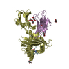
- PDB-6o9b: Crystal structure of HLA-A3*01 in complex with a wild-type beta-c... -
+ Open data

- Basic information
| Entry | Database: PDB / ID: 6o9b | |||||||||
|---|---|---|---|---|---|---|---|---|---|---|
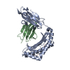
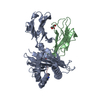
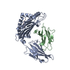
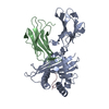
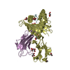
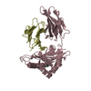
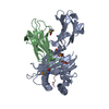
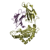
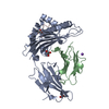
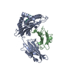
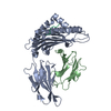
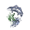
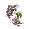
| Title | Crystal structure of HLA-A3*01 in complex with a wild-type beta-catenin peptide | |||||||||
 Components Components |
| |||||||||
 Keywords Keywords |  IMMUNE SYSTEM / HLA-A3 / MHC Class I IMMUNE SYSTEM / HLA-A3 / MHC Class I | |||||||||
| Function / homology |  Function and homology information Function and homology informationCDH11 homotypic and heterotypic interactions / Regulation of CDH19 Expression and Function / positive regulation of heparan sulfate proteoglycan biosynthetic process / lung induction / positive regulation of branching involved in lung morphogenesis / cranial ganglion development / renal vesicle formation / renal inner medulla development / renal outer medulla development / nephron tubule formation ...CDH11 homotypic and heterotypic interactions / Regulation of CDH19 Expression and Function / positive regulation of heparan sulfate proteoglycan biosynthetic process / lung induction / positive regulation of branching involved in lung morphogenesis / cranial ganglion development / renal vesicle formation / renal inner medulla development / renal outer medulla development / nephron tubule formation / mesenchymal stem cell differentiation / beta-catenin-ICAT complex / metanephros morphogenesis / genitalia morphogenesis / embryonic skeletal limb joint morphogenesis / neural plate development / glial cell fate determination / regulation of secondary heart field cardioblast proliferation / astrocyte-dopaminergic neuron signaling / negative regulation of mitotic cell cycle, embryonic / canonical Wnt signaling pathway involved in mesenchymal stem cell differentiation / oviduct development / beta-catenin-TCF7L2 complex / regulation of nephron tubule epithelial cell differentiation / negative regulation of mesenchymal to epithelial transition involved in metanephros morphogenesis / regulation of timing of anagen / Binding of TCF/LEF:CTNNB1 to target gene promoters / central nervous system vasculogenesis / RUNX3 regulates WNT signaling / regulation of centriole-centriole cohesion / Regulation of CDH11 function / regulation of centromeric sister chromatid cohesion / embryonic axis specification / endodermal cell fate commitment / regulation of fibroblast proliferation / Scrib-APC-beta-catenin complex / positive regulation of fibroblast growth factor receptor signaling pathway / beta-catenin-TCF complex / lens morphogenesis in camera-type eye / dorsal root ganglion development / synaptic vesicle clustering / acinar cell differentiation / dorsal/ventral axis specification / proximal/distal pattern formation / neuron fate determination / layer formation in cerebral cortex / positive regulation of myoblast proliferation / positive regulation of endothelial cell differentiation / sympathetic ganglion development / establishment of blood-retinal barrier / fungiform papilla formation / lung epithelial cell differentiation / embryonic foregut morphogenesis / hindbrain development / regulation of calcium ion import / positive regulation of determination of dorsal identity / positive regulation of skeletal muscle tissue development / ectoderm development / positive regulation of odontoblast differentiation / cranial skeletal system development / endothelial tube morphogenesis / regulation of protein localization to cell surface / hair cell differentiation / mesenchymal cell proliferation involved in lung development / detection of muscle stretch / smooth muscle cell differentiation / histone methyltransferase binding / midbrain dopaminergic neuron differentiation / presynaptic active zone cytoplasmic component / alpha-catenin binding / cellular response to indole-3-methanol / flotillin complex / Germ layer formation at gastrulation / establishment of blood-brain barrier / male genitalia development / negative regulation of oligodendrocyte differentiation / apicolateral plasma membrane / fascia adherens / epithelial cell proliferation involved in prostate gland development / embryonic brain development / Formation of definitive endoderm / epithelial cell differentiation involved in prostate gland development / positive regulation of epithelial cell proliferation involved in prostate gland development / regulation of smooth muscle cell proliferation / negative regulation of oxidative stress-induced neuron intrinsic apoptotic signaling pathway / oocyte development / beta-catenin destruction complex / lung-associated mesenchyme development / Formation of axial mesoderm / negative regulation of protein sumoylation / adherens junction assembly / embryonic heart tube development / Beta-catenin phosphorylation cascade / Signaling by GSK3beta mutants / CTNNB1 S33 mutants aren't phosphorylated / CTNNB1 S37 mutants aren't phosphorylated / CTNNB1 S45 mutants aren't phosphorylated / CTNNB1 T41 mutants aren't phosphorylated / Adherens junctions interactions / catenin complexSimilarity search - Function | |||||||||
| Biological species |  Homo sapiens (human) Homo sapiens (human) | |||||||||
| Method | X-RAY DIFFRACTION / SYNCHROTRON / MOLECULAR REPLACEMENT / molecular replacement / Resolution: 2.2 Å | |||||||||
 Authors Authors | Miller, M.S. / Gabelli, S.B. | |||||||||
| Funding support |  United States, 2items United States, 2items
| |||||||||
 Citation Citation | Journal: J.Biol.Chem. / Year: 2019 Title: An engineered antibody fragment targeting mutant beta-catenin via major histocompatibility complex I neoantigen presentation. Authors: Miller, M.S. / Douglass, J. / Hwang, M.S. / Skora, A.D. / Murphy, M. / Papadopoulos, N. / Kinzler, K.W. / Vogelstein, B. / Zhou, S. / Gabelli, S.B. | |||||||||
| History |
|
- Structure visualization
| Structure viewer | Molecule: MolmilJmol/JSmol |
|---|
- Downloads & links
-Download
| PDBx/mmCIF format | 6o9b.cif.gz | 105.6 KB | Display | PDBx/mmCIF format |
|---|---|---|---|---|
| PDB format | pdb6o9b.ent.gz | 77.2 KB | Display | PDB format |
| PDBx/mmJSON format | 6o9b.json.gz | Tree view | PDBx/mmJSON format | |
| Others |  Other downloads Other downloads |
-Validation report
| Arichive directory | https://data.pdbj.org/pub/pdb/validation_reports/o9/6o9bftp://data.pdbj.org/pub/pdb/validation_reports/o9/6o9b | HTTPS FTP |
|---|
-Related structure data
| Related structure data | 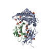 6o9cC 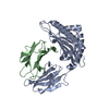 2xpgS S: Starting model for refinement C: citing same article ( |
|---|---|
| Similar structure data |


-Links
 PDBj
PDBj


- Assembly
| Deposited unit | 
| ||||||||
|---|---|---|---|---|---|---|---|---|---|
| 1 |
| ||||||||
| Unit cell |
|
-Components
-Protein , 2 types, 2 molecules AB
| #1: Protein | Mass: 34589.211 Da / Num. of mol.: 1 / Fragment: UNP residues 25-304 Source method: isolated from a genetically manipulated source Source: (gene. exp.) Homo sapiens (human) / Gene: HLA-A, HLAA / Production host:  Escherichia coli BL21(DE3) (bacteria) / References: UniProt: P04439 Escherichia coli BL21(DE3) (bacteria) / References: UniProt: P04439 |
|---|---|
| #2: Protein | Beta-2 microglobulin Mass: 13732.547 Da / Num. of mol.: 1 Source method: isolated from a genetically manipulated source Source: (gene. exp.) Homo sapiens (human) / Gene: B2M, CDABP0092, HDCMA22P / Production host: Escherichia coli BL21(DE3) (bacteria) / References: UniProt: P61769 |
-Protein/peptide , 1 types, 1 molecules C
| #3: Protein/peptide | / Beta-catenin Mass: 861.960 Da / Num. of mol.: 1 / Fragment: UNP residues 41-49 / Source method: obtained synthetically / Source: (synth.) Homo sapiens (human) / References: UniProt: P35222 |
|---|
-Non-polymers , 6 types, 184 molecules 


| #4: Chemical | Polyethylene glycol Mass: 194.226 Da / Num. of mol.: 2 / Source method: obtained synthetically / Formula: C8H18O5 / Comment: precipitant*YM Mass: 194.226 Da / Num. of mol.: 2 / Source method: obtained synthetically / Formula: C8H18O5 / Comment: precipitant*YM#5: Chemical | MES (buffer) Mass: 195.237 Da / Num. of mol.: 3 / Source method: obtained synthetically / Formula: C6H13NO4S / Comment: pH buffer*YM Mass: 195.237 Da / Num. of mol.: 3 / Source method: obtained synthetically / Formula: C6H13NO4S / Comment: pH buffer*YM#6: Chemical | Imidazole Mass: 69.085 Da / Num. of mol.: 2 / Source method: obtained synthetically / Formula: C3H5N2 Mass: 69.085 Da / Num. of mol.: 2 / Source method: obtained synthetically / Formula: C3H5N2#7: Chemical | ChemComp-PEG / Diethylene glycol Mass: 106.120 Da / Num. of mol.: 4 / Source method: obtained synthetically / Formula: C4H10O3 Mass: 106.120 Da / Num. of mol.: 4 / Source method: obtained synthetically / Formula: C4H10O3#8: Chemical | ChemComp-EDO / Ethylene glycol Mass: 62.068 Da / Num. of mol.: 7 / Source method: obtained synthetically / Formula: C2H6O2 Mass: 62.068 Da / Num. of mol.: 7 / Source method: obtained synthetically / Formula: C2H6O2#9: Water | ChemComp-HOH / | WaterMass: 18.015 Da / Num. of mol.: 166 / Source method: isolated from a natural source / Formula: H2O |
|---|
-Experimental details
-Experiment
| Experiment | Method: X-RAY DIFFRACTION / Number of used crystals: 1 |
|---|
- Sample preparation
| Crystal | Density Matthews: 2.91 Å3/Da / Density % sol: 57.66 % |
|---|---|
| Crystal grow | Temperature: 292 K / Method: vapor diffusion, hanging drop / pH: 6.5 Details: 0.1 M MES/imidazole, pH 6.5, 0.03 M diethylene glycol, 0.03 M triethylene glycol, 0.03 M tetraethylene glycol, 0.03 M pentaethylene glycol, 20% PEG500 MME, 10% PEG20000 |
-Data collection
| Diffraction | Mean temperature: 100 K / Serial crystal experiment: N | ||||||||||||||||||||||||
|---|---|---|---|---|---|---|---|---|---|---|---|---|---|---|---|---|---|---|---|---|---|---|---|---|---|
| Diffraction source | Source: SYNCHROTRON / Site: NSLS-II / Beamline: 17-ID-2 / Wavelength: 0.979 Å | ||||||||||||||||||||||||
| Detector | Type: DECTRIS EIGER X 16M / Detector: PIXEL / Date: Jun 23, 2017 | ||||||||||||||||||||||||
| Radiation | Monochromator: Si(111) / Protocol: SINGLE WAVELENGTH / Monochromatic (M) / Laue (L): M / Scattering type: x-ray | ||||||||||||||||||||||||
| Radiation wavelength | Wavelength: 0.979 Å / Relative weight: 1 | ||||||||||||||||||||||||
| Reflection | Resolution: 2.2→28.87 Å / Num. obs: 29999 / % possible obs: 99.2 % / Redundancy: 10.4 % / CC1/2: 0.996 / Rmerge(I) obs: 0.164 / Rpim(I) all: 0.052 / Rrim(I) all: 0.172 / Net I/σ(I): 9.7 / Num. measured all: 313372 | ||||||||||||||||||||||||
| Reflection shell | Diffraction-ID: 1
|
-Phasing
| Phasing | Method: molecular replacement | ||||||
|---|---|---|---|---|---|---|---|
| Phasing MR | R rigid body: 0.349
|
- Processing
| Software |
| ||||||||||||||||||||||||||||||||||||||||||||||||||||||||||||
|---|---|---|---|---|---|---|---|---|---|---|---|---|---|---|---|---|---|---|---|---|---|---|---|---|---|---|---|---|---|---|---|---|---|---|---|---|---|---|---|---|---|---|---|---|---|---|---|---|---|---|---|---|---|---|---|---|---|---|---|---|---|
| Refinement | Method to determine structure: MOLECULAR REPLACEMENT Starting model: PDB entry 2XPG Resolution: 2.2→28.87 Å / Cor.coef. Fo:Fc: 0.959 / Cor.coef. Fo:Fc free: 0.932 / SU B: 4.781 / SU ML: 0.123 / SU R Cruickshank DPI: 0.199 / Cross valid method: THROUGHOUT / σ(F): 0 / ESU R: 0.199 / ESU R Free: 0.177 Details: HYDROGENS HAVE BEEN ADDED IN THE RIDING POSITIONS U VALUES : REFINED INDIVIDUALLY
| ||||||||||||||||||||||||||||||||||||||||||||||||||||||||||||
| Solvent computation | Ion probe radii: 0.8 Å / Shrinkage radii: 0.8 Å / VDW probe radii: 1.2 Å | ||||||||||||||||||||||||||||||||||||||||||||||||||||||||||||
| Displacement parameters | Biso max: 120.15 Å2 / Biso mean: 37.94 Å2 / Biso min: 19.19 Å2
| ||||||||||||||||||||||||||||||||||||||||||||||||||||||||||||
| Refinement step | Cycle: final / Resolution: 2.2→28.87 Å
| ||||||||||||||||||||||||||||||||||||||||||||||||||||||||||||
| Refine LS restraints |
| ||||||||||||||||||||||||||||||||||||||||||||||||||||||||||||
| LS refinement shell | Resolution: 2.197→2.254 Å / Rfactor Rfree error: 0 / Total num. of bins used: 20
|
