 Movie
Movie Controller

[English] 日本語
 Yorodumi
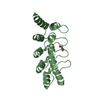
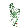
- PDB-6o2v: Crystal structure of the SARAF luminal domain Cys-lock mutant monomer -
+ Open data

- Basic information
| Entry | Database: PDB / ID: 6o2v | |||||||||||||||
|---|---|---|---|---|---|---|---|---|---|---|---|---|---|---|---|---|
| Title | Crystal structure of the SARAF luminal domain Cys-lock mutant monomer | |||||||||||||||
 Components Components | Store-operated calcium entry-associated regulatory factor | |||||||||||||||
 Keywords Keywords |  SIGNALING PROTEIN / SOCE / Store Operated Calcium Entry / SARAF / ER / Endoplasmic reticulum / Calcium signaling / Domain swap SIGNALING PROTEIN / SOCE / Store Operated Calcium Entry / SARAF / ER / Endoplasmic reticulum / Calcium signaling / Domain swap | |||||||||||||||
| Function / homology | Store-operated calcium entry-associated regulatory factor / SOCE-associated regulatory factor of calcium homoeostasis / endoplasmic reticulum-plasma membrane contact site / regulation of store-operated calcium entry / calcium ion transport / endoplasmic reticulum membrane / endoplasmic reticulum / FORMIC ACID / Store-operated calcium entry-associated regulatory factor Function and homology information Function and homology information | |||||||||||||||
| Biological species |  Homo sapiens (human) Homo sapiens (human) | |||||||||||||||
| Method | X-RAY DIFFRACTION / SYNCHROTRON / MOLECULAR REPLACEMENT / Resolution: 1.58 Å | |||||||||||||||
 Authors Authors | Kimberlin, C.R. / Minor, D.L. | |||||||||||||||
| Funding support |  United States, 4items United States, 4items
| |||||||||||||||
 Citation Citation | Journal: J.Mol.Biol. / Year: 2019 Title: SARAF Luminal Domain Structure Reveals a Novel Domain-Swapped beta-Sandwich Fold Important for SOCE Modulation. Authors: Kimberlin, C.R. / Meshcheriakova, A. / Palty, R. / Raveh, A. / Karbat, I. / Reuveny, E. / Minor Jr., D.L. | |||||||||||||||
| History |
|
- Structure visualization
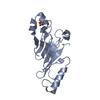
| Structure viewer | Molecule: MolmilJmol/JSmol |
|---|
- Downloads & links
-Download
| PDBx/mmCIF format | 6o2v.cif.gz | 78.1 KB | Display | PDBx/mmCIF format |
|---|---|---|---|---|
| PDB format | pdb6o2v.ent.gz | 56.1 KB | Display | PDB format |
| PDBx/mmJSON format | 6o2v.json.gz | Tree view | PDBx/mmJSON format | |
| Others |  Other downloads Other downloads |
-Validation report
| Arichive directory | https://data.pdbj.org/pub/pdb/validation_reports/o2/6o2vftp://data.pdbj.org/pub/pdb/validation_reports/o2/6o2v | HTTPS FTP |
|---|
-Related structure data
| Related structure data |  6o2uSC  6o2wC S: Starting model for refinement C: citing same article ( |
|---|---|
| Similar structure data |


-Links
 PDBj
PDBj- Assembly
| Deposited unit | 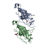
| ||||||||
|---|---|---|---|---|---|---|---|---|---|
| 1 | 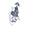
| ||||||||
| 2 | 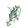
| ||||||||
| Unit cell |
|
-Components
| #1: Protein | Mass: 15523.334 Da / Num. of mol.: 2 / Mutation: K98C, A156C Source method: isolated from a genetically manipulated source Source: (gene. exp.) Homo sapiens (human)Gene: SARAF, TMEM66, XTP3, HSPC035, NPD003, PSEC0019, UNQ1967/PRO4499 Production host:  Escherichia coli (E. coli) / References: UniProt: Q96BY9 Escherichia coli (E. coli) / References: UniProt: Q96BY9#2: Chemical | ChemComp-FMT / | Formic acid  Mass: 46.025 Da / Num. of mol.: 1 / Source method: obtained synthetically / Formula: CH2O2 Mass: 46.025 Da / Num. of mol.: 1 / Source method: obtained synthetically / Formula: CH2O2#3: Chemical | ChemComp-GOL / | Glycerol  Mass: 92.094 Da / Num. of mol.: 1 / Source method: obtained synthetically / Formula: C3H8O3 Mass: 92.094 Da / Num. of mol.: 1 / Source method: obtained synthetically / Formula: C3H8O3#4: Water | ChemComp-HOH / | Water Mass: 18.015 Da / Num. of mol.: 306 / Source method: isolated from a natural source / Formula: H2O Mass: 18.015 Da / Num. of mol.: 306 / Source method: isolated from a natural source / Formula: H2O |
|---|
-Experimental details
-Experiment
| Experiment | Method: X-RAY DIFFRACTION / Number of used crystals: 1 |
|---|
- Sample preparation
| Crystal | Density Matthews: 1.86 Å3/Da / Density % sol: 33.82 % |
|---|---|
| Crystal grow | Temperature: 293 K / Method: vapor diffusion, hanging drop / pH: 4.4 Details: 0.1M sodium acetate, pH 4.2-4.6 and 1.2-1.6M sodium formate Temp details: room temperature |
-Data collection
| Diffraction | Mean temperature: 100 K / Serial crystal experiment: N | |||||||||||||||||||||||||||||||||||||||||||||||||||||||||||||||||||||||||||||||||||||||||||||||||||
|---|---|---|---|---|---|---|---|---|---|---|---|---|---|---|---|---|---|---|---|---|---|---|---|---|---|---|---|---|---|---|---|---|---|---|---|---|---|---|---|---|---|---|---|---|---|---|---|---|---|---|---|---|---|---|---|---|---|---|---|---|---|---|---|---|---|---|---|---|---|---|---|---|---|---|---|---|---|---|---|---|---|---|---|---|---|---|---|---|---|---|---|---|---|---|---|---|---|---|---|---|
| Diffraction source | Source: SYNCHROTRON / Site: ALS / Beamline: 8.3.1 / Wavelength: 1.116 Å | |||||||||||||||||||||||||||||||||||||||||||||||||||||||||||||||||||||||||||||||||||||||||||||||||||
| Detector | Type: ADSC QUANTUM 315r / Detector: CCD / Date: Apr 13, 2013 | |||||||||||||||||||||||||||||||||||||||||||||||||||||||||||||||||||||||||||||||||||||||||||||||||||
| Radiation | Protocol: SINGLE WAVELENGTH / Monochromatic (M) / Laue (L): M / Scattering type: x-ray | |||||||||||||||||||||||||||||||||||||||||||||||||||||||||||||||||||||||||||||||||||||||||||||||||||
| Radiation wavelength | Wavelength: 1.116 Å / Relative weight: 1 | |||||||||||||||||||||||||||||||||||||||||||||||||||||||||||||||||||||||||||||||||||||||||||||||||||
| Reflection | Resolution: 1.58→63.441 Å / Num. obs: 32028 / % possible obs: 98.7 % / Redundancy: 6.6 % / Biso Wilson estimate: 11.85 Å2 / Rpim(I) all: 0.038 / Rrim(I) all: 0.099 / Rsym value: 0.084 / Net I/av σ(I): 8.4 / Net I/σ(I): 16.2 | |||||||||||||||||||||||||||||||||||||||||||||||||||||||||||||||||||||||||||||||||||||||||||||||||||
| Reflection shell | Diffraction-ID: 1
|
- Processing
| Software |
| |||||||||||||||||||||||||||||||||||||||||||||||||||||||||||||||||||||||||||||||||||||||||||
|---|---|---|---|---|---|---|---|---|---|---|---|---|---|---|---|---|---|---|---|---|---|---|---|---|---|---|---|---|---|---|---|---|---|---|---|---|---|---|---|---|---|---|---|---|---|---|---|---|---|---|---|---|---|---|---|---|---|---|---|---|---|---|---|---|---|---|---|---|---|---|---|---|---|---|---|---|---|---|---|---|---|---|---|---|---|---|---|---|---|---|---|---|
| Refinement | Method to determine structure: MOLECULAR REPLACEMENT Starting model: 6O2U Resolution: 1.58→43.954 Å / SU ML: 0.15 / Cross valid method: THROUGHOUT / σ(F): 1.34 / Phase error: 19.64
| |||||||||||||||||||||||||||||||||||||||||||||||||||||||||||||||||||||||||||||||||||||||||||
| Solvent computation | Shrinkage radii: 0.9 Å / VDW probe radii: 1.11 Å | |||||||||||||||||||||||||||||||||||||||||||||||||||||||||||||||||||||||||||||||||||||||||||
| Displacement parameters | Biso max: 49.45 Å2 / Biso mean: 14.7208 Å2 / Biso min: 3.32 Å2 | |||||||||||||||||||||||||||||||||||||||||||||||||||||||||||||||||||||||||||||||||||||||||||
| Refinement step | Cycle: final / Resolution: 1.58→43.954 Å
| |||||||||||||||||||||||||||||||||||||||||||||||||||||||||||||||||||||||||||||||||||||||||||
| Refine LS restraints |
| |||||||||||||||||||||||||||||||||||||||||||||||||||||||||||||||||||||||||||||||||||||||||||
| LS refinement shell | Refine-ID: X-RAY DIFFRACTION / Rfactor Rfree error: 0 / Total num. of bins used: 12
|
