 Movie
Movie Controller

[English] 日本語
 Yorodumi
- PDB-6nor: Crystal structure of GenD2 from gentamicin A biosynthesis in comp... -
+ Open data

- Basic information
| Entry | Database: PDB / ID: 6nor | ||||||
|---|---|---|---|---|---|---|---|
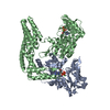
| Title | Crystal structure of GenD2 from gentamicin A biosynthesis in complex with NAD | ||||||
 Components Components | Putative NAD dependent dehydrogenase | ||||||
 Keywords Keywords |  OXIDOREDUCTASE / NAD depedent enzyme / 3D swapping domain OXIDOREDUCTASE / NAD depedent enzyme / 3D swapping domain | ||||||
| Function / homology | Gfo/Idh/MocA-like oxidoreductase, C-terminal / Oxidoreductase family, C-terminal alpha/beta domain / Gfo/Idh/MocA-like oxidoreductase, N-terminal / Oxidoreductase family, NAD-binding Rossmann fold / oxidoreductase activity / NAD(P)-binding domain superfamily / NICOTINAMIDE-ADENINE-DINUCLEOTIDE / Putative NAD dependent dehydrogenase Function and homology information Function and homology information | ||||||
| Biological species |  Micromonospora echinospora (bacteria) Micromonospora echinospora (bacteria) | ||||||
| Method | X-RAY DIFFRACTION / SYNCHROTRON / MOLECULAR REPLACEMENT / Resolution: 2.402 Å | ||||||
 Authors Authors | Araujo, N.C. / Bury, P.S. / Huang, F. / Leadlay, P.F. / Dias, M.V.B. | ||||||
 Citation Citation | Journal: Acs Chem.Biol. / Year: 2019 Title: Crystal Structure of GenD2, an NAD-Dependent Oxidoreductase Involved in the Biosynthesis of Gentamicin. Authors: de Araujo, N.C. / Bury, P.D.S. / Tavares, M.T. / Huang, F. / Parise-Filho, R. / Leadlay, P. / Dias, M.V.B. | ||||||
| History |
|
- Structure visualization
| Structure viewer | Molecule: MolmilJmol/JSmol |
|---|
- Downloads & links
-Download
| PDBx/mmCIF format | 6nor.cif.gz | 770 KB | Display | PDBx/mmCIF format |
|---|---|---|---|---|
| PDB format | pdb6nor.ent.gz | 642.2 KB | Display | PDB format |
| PDBx/mmJSON format | 6nor.json.gz | Tree view | PDBx/mmJSON format | |
| Others |  Other downloads Other downloads |
-Validation report
| Arichive directory | https://data.pdbj.org/pub/pdb/validation_reports/no/6norftp://data.pdbj.org/pub/pdb/validation_reports/no/6nor | HTTPS FTP |
|---|
-Related structure data
| Similar structure data |
|---|


-Links
 PDBj
PDBj- Assembly
| Deposited unit | 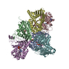
| |||||||||
|---|---|---|---|---|---|---|---|---|---|---|
| 1 | 
| |||||||||
| 2 | 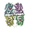
| |||||||||
| Unit cell |
| |||||||||
| Components on special symmetry positions |
|

-Components
| #1: Protein | Mass: 38719.457 Da / Num. of mol.: 6 Source method: isolated from a genetically manipulated source Source: (gene. exp.) Micromonospora echinospora (bacteria) / Gene: gtmC, genD2Production host: Escherichia coli 'BL21-Gold(DE3)pLysS AG' (bacteria)References: UniProt: Q70KD1 #2: Chemical | ChemComp-NAD / Nicotinamide adenine dinucleotide  Mass: 663.425 Da / Num. of mol.: 6 / Source method: obtained synthetically / Formula: C21H27N7O14P2 / Comment: NAD*YM Mass: 663.425 Da / Num. of mol.: 6 / Source method: obtained synthetically / Formula: C21H27N7O14P2 / Comment: NAD*YM#3: Water | ChemComp-HOH / | Water Mass: 18.015 Da / Num. of mol.: 436 / Source method: isolated from a natural source / Formula: H2O Mass: 18.015 Da / Num. of mol.: 436 / Source method: isolated from a natural source / Formula: H2O |
|---|
-Experimental details
-Experiment
| Experiment | Method: X-RAY DIFFRACTION / Number of used crystals: 1 |
|---|
- Sample preparation
| Crystal | Density Matthews: 2.71 Å3/Da / Density % sol: 54.69 % |
|---|---|
| Crystal grow | Temperature: 292 K / Method: vapor diffusion, hanging drop / pH: 4.6 / Details: 0.1 M sodium acetate, 4.6 M sodium formate |
-Data collection
| Diffraction | Mean temperature: 100 K / Serial crystal experiment: N | ||||||||||||||||||||||||
|---|---|---|---|---|---|---|---|---|---|---|---|---|---|---|---|---|---|---|---|---|---|---|---|---|---|
| Diffraction source | Source: SYNCHROTRON / Site: PETRA III, EMBL c/o DESY  / Beamline: P13 (MX1) / Wavelength: 0.97626 Å / Beamline: P13 (MX1) / Wavelength: 0.97626 Å | ||||||||||||||||||||||||
| Detector | Type: DECTRIS PILATUS 2M-F / Detector: PIXEL / Date: Dec 15, 2017 | ||||||||||||||||||||||||
| Radiation | Protocol: SINGLE WAVELENGTH / Monochromatic (M) / Laue (L): M / Scattering type: x-ray | ||||||||||||||||||||||||
| Radiation wavelength | Wavelength: 0.97626 Å / Relative weight: 1 | ||||||||||||||||||||||||
| Reflection | Resolution: 2.4→49.96 Å / Num. obs: 95469 / % possible obs: 100 % / Redundancy: 12.7 % / Biso Wilson estimate: 33.92 Å2 / CC1/2: 0.997 / Rmerge(I) obs: 0.179 / Rpim(I) all: 0.052 / Rrim(I) all: 0.187 / Net I/σ(I): 13.5 / Num. measured all: 1215767 / Scaling rejects: 36 | ||||||||||||||||||||||||
| Reflection shell | Diffraction-ID: 1
|
- Processing
| Software |
| |||||||||||||||||||||||||||||||||||||||||||||||||||||||||||||||||||||||||||||||||||||||||||||||
|---|---|---|---|---|---|---|---|---|---|---|---|---|---|---|---|---|---|---|---|---|---|---|---|---|---|---|---|---|---|---|---|---|---|---|---|---|---|---|---|---|---|---|---|---|---|---|---|---|---|---|---|---|---|---|---|---|---|---|---|---|---|---|---|---|---|---|---|---|---|---|---|---|---|---|---|---|---|---|---|---|---|---|---|---|---|---|---|---|---|---|---|---|---|---|---|---|
| Refinement | Method to determine structure: MOLECULAR REPLACEMENT / Resolution: 2.402→49.955 Å / SU ML: 0.25 / Cross valid method: THROUGHOUT / σ(F): 1.33 / Phase error: 22.28
| |||||||||||||||||||||||||||||||||||||||||||||||||||||||||||||||||||||||||||||||||||||||||||||||
| Solvent computation | Shrinkage radii: 0.9 Å / VDW probe radii: 1.11 Å | |||||||||||||||||||||||||||||||||||||||||||||||||||||||||||||||||||||||||||||||||||||||||||||||
| Displacement parameters | Biso max: 127.26 Å2 / Biso mean: 34.2915 Å2 / Biso min: 17.23 Å2 | |||||||||||||||||||||||||||||||||||||||||||||||||||||||||||||||||||||||||||||||||||||||||||||||
| Refinement step | Cycle: final / Resolution: 2.402→49.955 Å
| |||||||||||||||||||||||||||||||||||||||||||||||||||||||||||||||||||||||||||||||||||||||||||||||
| Refinement TLS params. | Method: refined / Origin x: 16.936 Å / Origin y: -39.331 Å / Origin z: -28.959 Å
| |||||||||||||||||||||||||||||||||||||||||||||||||||||||||||||||||||||||||||||||||||||||||||||||
| Refinement TLS group |
|
