- PDB-6njg: Ubiquitin Variant in Complex with Ubiquitin Interacting Motif -
+
Open data
ID or keywords:
Loading...
-
Basic information
Entry
Database: PDB / ID: 6njg
Title
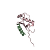
Ubiquitin Variant in Complex with Ubiquitin Interacting Motif
Components
Polyubiquitin-B
Vacuolar protein sorting-associated protein 27Vacuole
Keywords
PROTEIN BINDING / Ubiquitin Variant / UIM
Function / homology
Function and homology information
microlipophagy / positive regulation of protein maturation / ESCRT-0 complex / microautophagy / ATP export / protein retention in Golgi apparatus / protein transport to vacuole involved in ubiquitin-dependent protein catabolic process via the multivesicular body sorting pathway / protein targeting to vacuole / multivesicular body sorting pathway / late endosome to vacuole transport ...microlipophagy / positive regulation of protein maturation / ESCRT-0 complex / microautophagy / ATP export / protein retention in Golgi apparatus / protein transport to vacuole involved in ubiquitin-dependent protein catabolic process via the multivesicular body sorting pathway / protein targeting to vacuole / multivesicular body sorting pathway / late endosome to vacuole transport / ubiquitin-dependent protein catabolic process via the multivesicular body sorting pathway / phosphatidylinositol-3-phosphate binding / vacuolar membrane / protein secretion / ubiquitin binding / endosome membrane / endosome / protein heterodimerization activity / protein domain specific binding / protein-containing complex / metal ion binding / nucleus Similarity search - Function
In the structure databanks used in Yorodumi, some data are registered as the other names, "COVID-19 virus" and "2019-nCoV". Here are the details of the virus and the list of structure data.
Jan 31, 2019. EMDB accession codes are about to change! (news from PDBe EMDB page)
EMDB accession codes are about to change! (news from PDBe EMDB page)
The allocation of 4 digits for EMDB accession codes will soon come to an end. Whilst these codes will remain in use, new EMDB accession codes will include an additional digit and will expand incrementally as the available range of codes is exhausted. The current 4-digit format prefixed with “EMD-” (i.e. EMD-XXXX) will advance to a 5-digit format (i.e. EMD-XXXXX), and so on. It is currently estimated that the 4-digit codes will be depleted around Spring 2019, at which point the 5-digit format will come into force.
The EM Navigator/Yorodumi systems omit the EMD- prefix.
Related info.:Q: What is EMD? / ID/Accession-code notation in Yorodumi/EM Navigator
Yorodumi is a browser for structure data from EMDB, PDB, SASBDB, etc.
This page is also the successor to EM Navigator detail page, and also detail information page/front-end page for Omokage search.
The word "yorodu" (or yorozu) is an old Japanese word meaning "ten thousand". "mi" (miru) is to see.
Related info.:EMDB / PDB / SASBDB / Comparison of 3 databanks / Yorodumi Search / Aug 31, 2016. New EM Navigator & Yorodumi / Yorodumi Papers / Jmol/JSmol / Function and homology information / Changes in new EM Navigator and Yorodumi
 Movie
Movie Controller


 Open data
 Basic information
 Components
 Keywords
 PROTEIN BINDING / Ubiquitin Variant / UIM
 Function and homology information

 Authors
 Canada, 1items
  Citation
 Structure visualization
 Downloads & links
 Other downloads


 PDBj
PDBj


 Assembly

 Mass: 18.015 Da / Num. of mol.: 8 / Source method: isolated from a natural source / Formula: H2O
 Sample preparation
 Processing