 Movie
Movie Controller

[English] 日本語
 Yorodumi
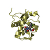
- PDB-6nh9: Crystal structure of a human calcium/calmodulin dependent serine ... -
+ Open data

- Basic information
| Entry | Database: PDB / ID: 6nh9 | ||||||
|---|---|---|---|---|---|---|---|
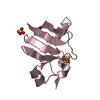
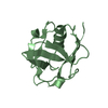
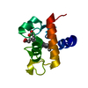
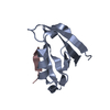
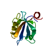
| Title | Crystal structure of a human calcium/calmodulin dependent serine protein kinase (CASK) PDZ domain | ||||||
 Components Components | Peripheral plasma membrane protein CASK | ||||||
 Keywords Keywords |  PROTEIN BINDING / PDZ domain / MAGUK protein family / peripheral plasma membrane protein / c-terminal peptide binding PROTEIN BINDING / PDZ domain / MAGUK protein family / peripheral plasma membrane protein / c-terminal peptide binding | ||||||
| Function / homology |  Function and homology information Function and homology informationnegative regulation of cellular response to growth factor stimulus / guanylate kinase activity / Dopamine Neurotransmitter Release Cycle / neurexin family protein binding / regulation of neurotransmitter secretion / negative regulation of wound healing / nuclear lamina / calcium ion import / Assembly and cell surface presentation of NMDA receptors / Neurexins and neuroligins ...negative regulation of cellular response to growth factor stimulus / guanylate kinase activity / Dopamine Neurotransmitter Release Cycle / neurexin family protein binding / regulation of neurotransmitter secretion / negative regulation of wound healing / nuclear lamina / calcium ion import / Assembly and cell surface presentation of NMDA receptors / Neurexins and neuroligins / Sensory processing of sound by outer hair cells of the cochlea / Sensory processing of sound by inner hair cells of the cochlea / Nephrin family interactions / ciliary membrane / regulation of synaptic vesicle exocytosis / Syndecan interactions / negative regulation of cell-matrix adhesion / positive regulation of calcium ion import / basement membrane / negative regulation of keratinocyte proliferation / establishment of localization in cell / Schaffer collateral - CA1 synapse / nuclear matrix / cell-cell junction / actin cytoskeleton / presynaptic membrane / basolateral plasma membrane / vesicle / calmodulin binding / non-specific serine/threonine protein kinase / cell adhesion / phosphorylation / signaling receptor binding / focal adhesion / protein serine kinase activity / protein serine/threonine kinase activity / nucleolus / positive regulation of transcription by RNA polymerase II / ATP binding / plasma membrane / cytosol / cytoplasmSimilarity search - Function | ||||||
| Biological species |  Homo sapiens (human) Homo sapiens (human) | ||||||
| Method | X-RAY DIFFRACTION / SYNCHROTRON / Resolution: 1.85 Å | ||||||
 Authors Authors | Sun, Y.J. / Gakhar, L. / Fuentes, E.J. | ||||||
| Funding support |  United States, 1items United States, 1items
| ||||||
 Citation Citation | Journal: To be published Title: CASK PDZ domain specificity Authors: Sun, Y.J. / Hou, T. / Fuentes, E.J. | ||||||
| History |
|
- Structure visualization
| Structure viewer | Molecule: MolmilJmol/JSmol |
|---|
- Downloads & links
-Download
| PDBx/mmCIF format | 6nh9.cif.gz | 162.6 KB | Display | PDBx/mmCIF format |
|---|---|---|---|---|
| PDB format | pdb6nh9.ent.gz | 132 KB | Display | PDB format |
| PDBx/mmJSON format | 6nh9.json.gz | Tree view | PDBx/mmJSON format | |
| Others |  Other downloads Other downloads |
-Validation report
| Arichive directory | https://data.pdbj.org/pub/pdb/validation_reports/nh/6nh9ftp://data.pdbj.org/pub/pdb/validation_reports/nh/6nh9 | HTTPS FTP |
|---|
-Related structure data


-Links
 PDBj
PDBj


- Assembly
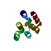
| Deposited unit | 
| ||||||||
|---|---|---|---|---|---|---|---|---|---|
| 1 | 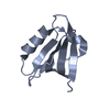
| ||||||||
| 2 | 
| ||||||||
| 3 | 
| ||||||||
| Unit cell |
| ||||||||
| Components on special symmetry positions |
|
-Components
| #1: Protein | Mass: 10118.886 Da / Num. of mol.: 3 Source method: isolated from a genetically manipulated source Details: M 485 Expression artifact G 486 Expression artifact Source: (gene. exp.) Homo sapiens (human) / Gene: CASK, LIN2 / Production host:  Escherichia coli (E. coli) Escherichia coli (E. coli)References: UniProt: O14936, non-specific serine/threonine protein kinase#2: Water | ChemComp-HOH / | Water Mass: 18.015 Da / Num. of mol.: 151 / Source method: isolated from a natural source / Formula: H2O Mass: 18.015 Da / Num. of mol.: 151 / Source method: isolated from a natural source / Formula: H2O |
|---|
-Experimental details
-Experiment
| Experiment | Method: X-RAY DIFFRACTION / Number of used crystals: 1 |
|---|
- Sample preparation
| Crystal | Density Matthews: 2.13 Å3/Da / Density % sol: 42.16 % |
|---|---|
| Crystal grow | Temperature: 291 K / Method: vapor diffusion, hanging drop / pH: 10.5 / Details: 30% v/v PEG 400 0.1M CAPS |
-Data collection
| Diffraction | Mean temperature: 100 K / Serial crystal experiment: N |
|---|---|
| Diffraction source | Source: SYNCHROTRON / Site: ALS / Beamline: 4.2.2 / Wavelength: 1.0003 Å |
| Detector | Type: RDI CMOS_8M / Detector: CMOS / Date: Sep 23, 2014 |
| Radiation | Monochromator: Si(111) / Protocol: SINGLE WAVELENGTH / Monochromatic (M) / Laue (L): M / Scattering type: x-ray |
| Radiation wavelength | Wavelength: 1.0003 Å / Relative weight: 1 |
| Reflection | Resolution: 1.85→59.77 Å / Num. obs: 20769 / % possible obs: 93.7 % / Redundancy: 1.8 % / CC1/2: 0.998 / Rmerge(I) obs: 0.021 / Rpim(I) all: 0.021 / Rrim(I) all: 0.03 / Net I/σ(I): 10.4 |
| Reflection shell | Resolution: 1.85→1.92 Å / Redundancy: 1.7 % / Rmerge(I) obs: 0.284 / Mean I/σ(I) obs: 2.1 / Num. unique obs: 1910 / CC1/2: 0.911 / Rpim(I) all: 0.284 / Rrim(I) all: 0.402 / % possible all: 93.7 |
- Processing
| Software |
| ||||||||||||||||||||||||||||||||||||||||||||||||||||||||
|---|---|---|---|---|---|---|---|---|---|---|---|---|---|---|---|---|---|---|---|---|---|---|---|---|---|---|---|---|---|---|---|---|---|---|---|---|---|---|---|---|---|---|---|---|---|---|---|---|---|---|---|---|---|---|---|---|---|
| Refinement | Resolution: 1.85→59.743 Å / SU ML: 0.28 / Cross valid method: FREE R-VALUE / σ(F): 1.34 / Phase error: 33.65
| ||||||||||||||||||||||||||||||||||||||||||||||||||||||||
| Solvent computation | Shrinkage radii: 0.9 Å / VDW probe radii: 1.11 Å | ||||||||||||||||||||||||||||||||||||||||||||||||||||||||
| Refinement step | Cycle: LAST / Resolution: 1.85→59.743 Å
| ||||||||||||||||||||||||||||||||||||||||||||||||||||||||
| Refine LS restraints |
| ||||||||||||||||||||||||||||||||||||||||||||||||||||||||
| LS refinement shell |
| ||||||||||||||||||||||||||||||||||||||||||||||||||||||||
| Refinement TLS params. | Method: refined / Origin x: 12.001 Å / Origin y: 5.2988 Å / Origin z: 30.4494 Å
| ||||||||||||||||||||||||||||||||||||||||||||||||||||||||
| Refinement TLS group | Selection details: all |
