 Movie
Movie Controller

[English] 日本語
 Yorodumi
- PDB-6nfu: Structure of the KcsA-G77A mutant or the 2,4-ion bound configurat... -
+ Open data

- Basic information
| Entry | Database: PDB / ID: 6nfu | |||||||||||||||
|---|---|---|---|---|---|---|---|---|---|---|---|---|---|---|---|---|
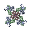
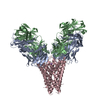
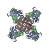
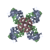
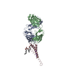
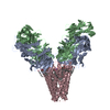
| Title | Structure of the KcsA-G77A mutant or the 2,4-ion bound configuration of a K+ channel selectivity filter. | |||||||||||||||
 Components Components |
| |||||||||||||||
 Keywords Keywords |  membrane protein / metal transport / ion channel / membrane transport / potassium channel membrane protein / metal transport / ion channel / membrane transport / potassium channel | |||||||||||||||
| Function / homology |  Function and homology information Function and homology informationdelayed rectifier potassium channel activity / voltage-gated potassium channel complex / identical protein bindingSimilarity search - Function | |||||||||||||||
| Biological species |  Mus musculus (house mouse) Mus musculus (house mouse) Streptomyces lividans (bacteria) Streptomyces lividans (bacteria) | |||||||||||||||
| Method | X-RAY DIFFRACTION / SYNCHROTRON / MOLECULAR REPLACEMENT / Resolution: 2.09 Å | |||||||||||||||
 Authors Authors | Tilegenova, C. / Cortes, D.M. / Jahovic, N. / Hardy, E. / Parameswaran, H. / Guan, L. / Cuello, L.G. | |||||||||||||||
| Funding support |  United States, 4items United States, 4items
| |||||||||||||||
 Citation Citation | Journal: Proc.Natl.Acad.Sci.USA / Year: 2019 Title: Structure, function, and ion-binding properties of a K+channel stabilized in the 2,4-ion-bound configuration. Authors: Tilegenova, C. / Cortes, D.M. / Jahovic, N. / Hardy, E. / Hariharan, P. / Guan, L. / Cuello, L.G. | |||||||||||||||
| History |
|
- Structure visualization
| Structure viewer | Molecule: MolmilJmol/JSmol |
|---|
- Downloads & links
-Download
| PDBx/mmCIF format | 6nfu.cif.gz | 127.2 KB | Display | PDBx/mmCIF format |
|---|---|---|---|---|
| PDB format | pdb6nfu.ent.gz | 94 KB | Display | PDB format |
| PDBx/mmJSON format | 6nfu.json.gz | Tree view | PDBx/mmJSON format | |
| Others |  Other downloads Other downloads |
-Validation report
| Arichive directory | https://data.pdbj.org/pub/pdb/validation_reports/nf/6nfuftp://data.pdbj.org/pub/pdb/validation_reports/nf/6nfu | HTTPS FTP |
|---|
-Related structure data
| Related structure data |  6nfvC  6pa0C  1k4cS S: Starting model for refinement C: citing same article ( |
|---|---|
| Similar structure data |


-Links
 PDBj
PDBj


- Assembly
| Deposited unit | 
| ||||||||||||||||||||||||||||||
|---|---|---|---|---|---|---|---|---|---|---|---|---|---|---|---|---|---|---|---|---|---|---|---|---|---|---|---|---|---|---|---|
| 1 | 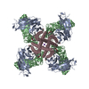
| ||||||||||||||||||||||||||||||
| Unit cell |
| ||||||||||||||||||||||||||||||
| Components on special symmetry positions |
|
-Components
-Protein , 1 types, 1 molecules C
| #3: Protein | Mass: 10992.763 Da / Num. of mol.: 1 / Mutation: G77C, L90C Source method: isolated from a genetically manipulated source Source: (gene. exp.) Streptomyces lividans (bacteria) / Gene: kcsA, skc1 / Production host: Escherichia coli (E. coli) / References: UniProt: P0A334 |
|---|
-Antibody , 2 types, 2 molecules AB
| #1: Antibody | Mass: 23411.242 Da / Num. of mol.: 1 Source method: isolated from a genetically manipulated source Source: (gene. exp.) Mus musculus (house mouse) / Production host: Mammalia (mammals) |
|---|---|
| #2: Antibody | Mass: 23435.738 Da / Num. of mol.: 1 Source method: isolated from a genetically manipulated source Source: (gene. exp.) Mus musculus (house mouse) / Production host: Mammalia (mammals) |
-Non-polymers , 4 types, 311 molecules 


| #4: Chemical | ChemComp-F09 / 1-Nonanol Mass: 144.254 Da / Num. of mol.: 1 / Source method: obtained synthetically / Formula: C9H20O Mass: 144.254 Da / Num. of mol.: 1 / Source method: obtained synthetically / Formula: C9H20O | ||
|---|---|---|---|
| #5: Chemical | ChemComp-1EM / ( Mass: 442.672 Da / Num. of mol.: 1 / Source method: obtained synthetically / Formula: C26H50O5 Mass: 442.672 Da / Num. of mol.: 1 / Source method: obtained synthetically / Formula: C26H50O5 | ||
| #6: Chemical | ChemComp-K /  Mass: 39.098 Da / Num. of mol.: 4 / Source method: obtained synthetically / Formula: K Mass: 39.098 Da / Num. of mol.: 4 / Source method: obtained synthetically / Formula: K#7: Water | ChemComp-HOH / | WaterMass: 18.015 Da / Num. of mol.: 305 / Source method: isolated from a natural source / Formula: H2O |
-Experimental details
-Experiment
| Experiment | Method: X-RAY DIFFRACTION / Number of used crystals: 1 |
|---|
- Sample preparation
| Crystal | Density Matthews: 3.94 Å3/Da / Density % sol: 68.78 % |
|---|---|
| Crystal grow | Temperature: 293 K / Method: vapor diffusion, sitting drop / Details: PEG400, magnesium acetate, sodium acetate |
-Data collection
| Diffraction | Mean temperature: 100 K / Serial crystal experiment: N |
|---|---|
| Diffraction source | Source: SYNCHROTRON / Site: SSRL / Beamline: BL14-1 / Wavelength: 0.987 Å |
| Detector | Type: MARMOSAIC 325 mm CCD / Detector: CCD / Date: Nov 25, 2015 |
| Radiation | Protocol: SINGLE WAVELENGTH / Monochromatic (M) / Laue (L): M / Scattering type: x-ray |
| Radiation wavelength | Wavelength: 0.987 Å / Relative weight: 1 |
| Reflection | Resolution: 2.09→34.678 Å / Num. obs: 53355 / % possible obs: 99.89 % / Redundancy: 7.9 % / Biso Wilson estimate: 35.46 Å2 / Net I/σ(I): 30.38 |
| Reflection shell | Resolution: 2.09→34.678 Å |
- Processing
| Software |
| ||||||||||||||||||||||||||||||||||||||||||||||||||||||||||||
|---|---|---|---|---|---|---|---|---|---|---|---|---|---|---|---|---|---|---|---|---|---|---|---|---|---|---|---|---|---|---|---|---|---|---|---|---|---|---|---|---|---|---|---|---|---|---|---|---|---|---|---|---|---|---|---|---|---|---|---|---|---|
| Refinement | Method to determine structure: MOLECULAR REPLACEMENT Starting model: 1k4c Resolution: 2.09→34.678 Å / SU ML: 0.2 / Cross valid method: THROUGHOUT / σ(F): 1.34 / Phase error: 24.85
| ||||||||||||||||||||||||||||||||||||||||||||||||||||||||||||
| Solvent computation | Shrinkage radii: 0.9 Å / VDW probe radii: 1.11 Å | ||||||||||||||||||||||||||||||||||||||||||||||||||||||||||||
| Displacement parameters | Biso max: 85.01 Å2 / Biso mean: 43.4086 Å2 / Biso min: 19.23 Å2 | ||||||||||||||||||||||||||||||||||||||||||||||||||||||||||||
| Refinement step | Cycle: final / Resolution: 2.09→34.678 Å
| ||||||||||||||||||||||||||||||||||||||||||||||||||||||||||||
| LS refinement shell | Refine-ID: X-RAY DIFFRACTION / Rfactor Rfree error: 0 / Total num. of bins used: 9 / % reflection obs: 100 %
|
