 Movie
Movie Controller

[English] 日本語
 Yorodumi
- PDB-6n9m: Crystal Structure of Adenosine Deaminase from Salmonella typhimur... -
+ Open data

- Basic information
| Entry | Database: PDB / ID: 6n9m | ||||||
|---|---|---|---|---|---|---|---|
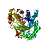
| Title | Crystal Structure of Adenosine Deaminase from Salmonella typhimurium with Pentostatin (Deoxycoformycin) | ||||||
 Components Components | Adenosine deaminase | ||||||
 Keywords Keywords | HYDROLASE / deaminase / pentostatin / Structural Genomics / Center for Structural Genomics of Infectious Diseases / CSGID | ||||||
| Function / homology |  Function and homology information Function and homology informationinosine biosynthetic process / adenosine deaminase / adenosine catabolic process / adenosine deaminase activity / hypoxanthine salvage / purine ribonucleoside monophosphate biosynthetic process / nucleotide metabolic process / zinc ion binding / cytosolSimilarity search - Function | ||||||
| Biological species |  Salmonella typhimurium (bacteria) Salmonella typhimurium (bacteria) | ||||||
| Method | X-RAY DIFFRACTION / SYNCHROTRON / SAD / Resolution: 1.449 Å | ||||||
 Authors Authors | Maltseva, N. / Kim, Y. / Grimshaw, S. / Joachimiak, A. / Center for Structural Genomics of Infectious Diseases (CSGID) | ||||||
 Citation Citation | Journal: To Be Published Title: Crystal Structure of Adenosine Deaminase from Salmonella typhimurium Complexed with Pentostatin (Deoxycoformycin) (CASP target) Authors: Maltseva, N. / Kim, Y. / Grimshaw, S. / Joachimiak, A. | ||||||
| History |
|
- Structure visualization
| Structure viewer | Molecule: MolmilJmol/JSmol |
|---|
- Downloads & links
-Download
| PDBx/mmCIF format | 6n9m.cif.gz | 83.9 KB | Display | PDBx/mmCIF format |
|---|---|---|---|---|
| PDB format | pdb6n9m.ent.gz | 66.2 KB | Display | PDB format |
| PDBx/mmJSON format | 6n9m.json.gz | Tree view | PDBx/mmJSON format | |
| Others |  Other downloads Other downloads |
-Validation report
| Arichive directory | https://data.pdbj.org/pub/pdb/validation_reports/n9/6n9mftp://data.pdbj.org/pub/pdb/validation_reports/n9/6n9m | HTTPS FTP |
|---|
-Related structure data
| Similar structure data | |
|---|---|
| Other databases |


-Links
 PDBj
PDBj

- Assembly
| Deposited unit | 
| ||||||||
|---|---|---|---|---|---|---|---|---|---|
| 1 |
| ||||||||
| Unit cell |
|
-Components
-Protein , 1 types, 1 molecules A
| #1: Protein | / Adenosine aminohydrolase Mass: 37520.672 Da / Num. of mol.: 1 Source method: isolated from a genetically manipulated source Source: (gene. exp.) Salmonella typhimurium (strain LT2 / SGSC1412 / ATCC 700720) (bacteria)Strain: LT2 / SGSC1412 / ATCC 700720 / Gene: add, STM1463 / Plasmid: pMCSG28 / Production host: Escherichia coli BL21(DE3) (bacteria) / Strain (production host): BL21-Gold (DE3) / References: UniProt: Q8ZPL9, adenosine deaminase |
|---|
-Non-polymers , 5 types, 211 molecules 


| #2: Chemical | ChemComp-DCF / Pentostatin Mass: 268.269 Da / Num. of mol.: 1 / Source method: obtained synthetically / Formula: C11H16N4O4 / Comment: anticancer*YM Mass: 268.269 Da / Num. of mol.: 1 / Source method: obtained synthetically / Formula: C11H16N4O4 / Comment: anticancer*YM | ||||
|---|---|---|---|---|---|
| #3: Chemical | ChemComp-ZN /  Mass: 65.409 Da / Num. of mol.: 1 / Source method: obtained synthetically / Formula: Zn Mass: 65.409 Da / Num. of mol.: 1 / Source method: obtained synthetically / Formula: Zn | ||||
| #4: Chemical |  Mass: 40.078 Da / Num. of mol.: 2 / Source method: obtained synthetically / Formula: Ca Mass: 40.078 Da / Num. of mol.: 2 / Source method: obtained synthetically / Formula: Ca#5: Chemical | ChemComp-FMT / | Formic acid Mass: 46.025 Da / Num. of mol.: 1 / Source method: obtained synthetically / Formula: CH2O2 Mass: 46.025 Da / Num. of mol.: 1 / Source method: obtained synthetically / Formula: CH2O2#6: Water | ChemComp-HOH / | WaterMass: 18.015 Da / Num. of mol.: 206 / Source method: isolated from a natural source / Formula: H2O |
-Experimental details
-Experiment
| Experiment | Method: X-RAY DIFFRACTION / Number of used crystals: 1 |
|---|
- Sample preparation
| Crystal | Density Matthews: 1.9 Å3/Da / Density % sol: 35.21 % |
|---|---|
| Crystal grow | Temperature: 289 K / Method: vapor diffusion, sitting drop / Details: 20% PEG 3000, 0.1M Tris pH7.0, 0.2M Ca(OAc) |
-Data collection
| Diffraction | Mean temperature: 100 K / Serial crystal experiment: N |
|---|---|
| Diffraction source | Source: SYNCHROTRON / Site: APS  / Beamline: 19-ID / Wavelength: 0.97918 Å / Beamline: 19-ID / Wavelength: 0.97918 Å |
| Detector | Type: DECTRIS PILATUS3 S 6M / Detector: PIXEL / Date: Dec 17, 2017 |
| Radiation | Monochromator: Si(111) / Protocol: SINGLE WAVELENGTH / Monochromatic (M) / Laue (L): M / Scattering type: x-ray |
| Radiation wavelength | Wavelength: 0.97918 Å / Relative weight: 1 |
| Reflection | Resolution: 1.449→50 Å / Num. obs: 50915 / % possible obs: 99.3 % / Redundancy: 5.8 % / Biso Wilson estimate: 17.4 Å2 / CC1/2: 0.715 / Rsym value: 0.098 / Net I/σ(I): 23.7 |
| Reflection shell | Resolution: 1.45→1.48 Å / Redundancy: 4.5 % / Mean I/σ(I) obs: 2.4 / Num. unique obs: 2471 / Rsym value: 0.683 / % possible all: 99.4 |
- Processing
| Software |
| |||||||||||||||||||||||||||||||||||||||||||||||||||||||||||||||||||||||||||||||||||||||||||||||||||||||||||||||||||||||||||||||||||||
|---|---|---|---|---|---|---|---|---|---|---|---|---|---|---|---|---|---|---|---|---|---|---|---|---|---|---|---|---|---|---|---|---|---|---|---|---|---|---|---|---|---|---|---|---|---|---|---|---|---|---|---|---|---|---|---|---|---|---|---|---|---|---|---|---|---|---|---|---|---|---|---|---|---|---|---|---|---|---|---|---|---|---|---|---|---|---|---|---|---|---|---|---|---|---|---|---|---|---|---|---|---|---|---|---|---|---|---|---|---|---|---|---|---|---|---|---|---|---|---|---|---|---|---|---|---|---|---|---|---|---|---|---|---|---|
| Refinement | Method to determine structure: SAD / Resolution: 1.449→45.937 Å / SU ML: 0.12 / Cross valid method: THROUGHOUT / σ(F): 0.31 / Phase error: 14.44
| |||||||||||||||||||||||||||||||||||||||||||||||||||||||||||||||||||||||||||||||||||||||||||||||||||||||||||||||||||||||||||||||||||||
| Solvent computation | Shrinkage radii: 0.9 Å / VDW probe radii: 1.11 Å | |||||||||||||||||||||||||||||||||||||||||||||||||||||||||||||||||||||||||||||||||||||||||||||||||||||||||||||||||||||||||||||||||||||
| Refinement step | Cycle: LAST / Resolution: 1.449→45.937 Å
| |||||||||||||||||||||||||||||||||||||||||||||||||||||||||||||||||||||||||||||||||||||||||||||||||||||||||||||||||||||||||||||||||||||
| Refine LS restraints |
| |||||||||||||||||||||||||||||||||||||||||||||||||||||||||||||||||||||||||||||||||||||||||||||||||||||||||||||||||||||||||||||||||||||
| LS refinement shell |
|
