 Movie
Movie Controller


+ Open data

- Basic information
| Entry | Database: PDB / ID: 6n3r | ||||||
|---|---|---|---|---|---|---|---|
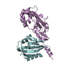
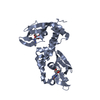
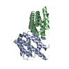
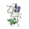
| Title | Sheep Galectin-11 (LGALS11) complex with galactose | ||||||
 Components Components | Galectin | ||||||
 Keywords Keywords | SUGAR BINDING PROTEIN / Glycan binding / Galectin / Lectin | ||||||
| Function / homology |  Function and homology information Function and homology information | ||||||
| Biological species |  Ovis aries (sheep) Ovis aries (sheep) | ||||||
| Method | X-RAY DIFFRACTION / SYNCHROTRON / MOLECULAR REPLACEMENT / molecular replacement / Resolution: 2.397 Å | ||||||
 Authors Authors | Beddoe, T.C. / Sakthivel, D. | ||||||
 Citation Citation | Journal: To be Published Title: Sheep Galectin-11 (LGALS11) complex with galactose Authors: Beddoe, T.C. / Sakthivel, D. #1: Journal: Acta Crystallogr F Struct Biol Commun / Year: 2015 Title: Cloning, expression, purification and crystallographic studies of galectin-11 from domestic sheep (Ovis aries). Authors: Sakthivel, D. / Littler, D. / Shahine, A. / Troy, S. / Johnson, M. / Rossjohn, J. / Piedrafita, D. / Beddoe, T. | ||||||
| History |
|
- Structure visualization
| Structure viewer | Molecule: MolmilJmol/JSmol |
|---|
- Downloads & links
-Download
| PDBx/mmCIF format | 6n3r.cif.gz | 243.1 KB | Display | PDBx/mmCIF format |
|---|---|---|---|---|
| PDB format | pdb6n3r.ent.gz | 195.3 KB | Display | PDB format |
| PDBx/mmJSON format | 6n3r.json.gz | Tree view | PDBx/mmJSON format | |
| Others |  Other downloads Other downloads |
-Validation report
| Arichive directory | https://data.pdbj.org/pub/pdb/validation_reports/n3/6n3rftp://data.pdbj.org/pub/pdb/validation_reports/n3/6n3r | HTTPS FTP |
|---|
-Related structure data
| Similar structure data |
|---|


-Links
 PDBj
PDBj

- Assembly
| Deposited unit | 
| ||||||||
|---|---|---|---|---|---|---|---|---|---|
| 1 | 
| ||||||||
| 2 | 
| ||||||||
| 3 | 
| ||||||||
| 4 | 
| ||||||||
| Unit cell |
|
-Components
| #1: Protein | / Galectin-11 Mass: 15580.910 Da / Num. of mol.: 8 Source method: isolated from a genetically manipulated source Source: (gene. exp.) Ovis aries (sheep) / Strain: Merino / Plasmid: pET28 / Production host:  Escherichia coli BL21 (bacteria) / References: UniProt: A0A0A7EMW6 Escherichia coli BL21 (bacteria) / References: UniProt: A0A0A7EMW6#2: Sugar | ChemComp-GAL / Galactose  Type: D-saccharide, beta linking / Mass: 180.156 Da / Num. of mol.: 5 Type: D-saccharide, beta linking / Mass: 180.156 Da / Num. of mol.: 5Source method: isolated from a genetically manipulated source Formula: C6H12O6 #3: Water | ChemComp-HOH / | Water Mass: 18.015 Da / Num. of mol.: 1064 / Source method: isolated from a natural source / Formula: H2O Mass: 18.015 Da / Num. of mol.: 1064 / Source method: isolated from a natural source / Formula: H2O |
|---|
-Experimental details
-Experiment
| Experiment | Method: X-RAY DIFFRACTION / Number of used crystals: 1 |
|---|
- Sample preparation
| Crystal | Density Matthews: 3.45 Å3/Da / Density % sol: 64.32 % |
|---|---|
| Crystal grow | Temperature: 293 K / Method: vapor diffusion, hanging drop / pH: 7.5 Details: 2% v/v Tacsimate, pH 7.0, 0.1 M HEPES, pH 7.5, 20% w/v PEG3350 PH range: 7.3 - 7.8 |
-Data collection
| Diffraction | Mean temperature: 100 K / Serial crystal experiment: N |
|---|---|
| Diffraction source | Source: SYNCHROTRON / Site: Australian Synchrotron  / Beamline: MX1 / Wavelength: 0.9537 Å / Beamline: MX1 / Wavelength: 0.9537 Å |
| Detector | Type: ADSC QUANTUM 210r / Detector: CCD / Date: Aug 6, 2014 |
| Radiation | Monochromator: double crystal Si(111) / Protocol: SINGLE WAVELENGTH / Monochromatic (M) / Laue (L): M / Scattering type: x-ray |
| Radiation wavelength | Wavelength: 0.9537 Å / Relative weight: 1 |
| Reflection | Resolution: 2.397→47.169 Å / Num. obs: 68286 / % possible obs: 99.97 % / Redundancy: 7.9 % / Biso Wilson estimate: 22.3 Å2 / Net I/σ(I): 7.3 |
| Reflection shell | Resolution: 2.71→2.81 Å |
-Phasing
| Phasing | Method: molecular replacement |
|---|
- Processing
| Software |
| ||||||||||||||||||||||||||||||||||||||||||||||||||||||||||||||||||||||||||||||||||||||||||||||||||||||||||||||||||||||||||||||||||||||||||||||||||||||||||||||||||||||||||||||||||||||
|---|---|---|---|---|---|---|---|---|---|---|---|---|---|---|---|---|---|---|---|---|---|---|---|---|---|---|---|---|---|---|---|---|---|---|---|---|---|---|---|---|---|---|---|---|---|---|---|---|---|---|---|---|---|---|---|---|---|---|---|---|---|---|---|---|---|---|---|---|---|---|---|---|---|---|---|---|---|---|---|---|---|---|---|---|---|---|---|---|---|---|---|---|---|---|---|---|---|---|---|---|---|---|---|---|---|---|---|---|---|---|---|---|---|---|---|---|---|---|---|---|---|---|---|---|---|---|---|---|---|---|---|---|---|---|---|---|---|---|---|---|---|---|---|---|---|---|---|---|---|---|---|---|---|---|---|---|---|---|---|---|---|---|---|---|---|---|---|---|---|---|---|---|---|---|---|---|---|---|---|---|---|---|---|
| Refinement | Method to determine structure: MOLECULAR REPLACEMENT / Resolution: 2.397→47.169 Å / SU ML: 0.24 / Cross valid method: THROUGHOUT / σ(F): 1.34 / Phase error: 19.73 / Stereochemistry target values: ML
| ||||||||||||||||||||||||||||||||||||||||||||||||||||||||||||||||||||||||||||||||||||||||||||||||||||||||||||||||||||||||||||||||||||||||||||||||||||||||||||||||||||||||||||||||||||||
| Solvent computation | Shrinkage radii: 0.9 Å / VDW probe radii: 1.11 Å / Solvent model: FLAT BULK SOLVENT MODEL | ||||||||||||||||||||||||||||||||||||||||||||||||||||||||||||||||||||||||||||||||||||||||||||||||||||||||||||||||||||||||||||||||||||||||||||||||||||||||||||||||||||||||||||||||||||||
| Displacement parameters | Biso max: 79.18 Å2 / Biso mean: 24.3736 Å2 / Biso min: 7.79 Å2 | ||||||||||||||||||||||||||||||||||||||||||||||||||||||||||||||||||||||||||||||||||||||||||||||||||||||||||||||||||||||||||||||||||||||||||||||||||||||||||||||||||||||||||||||||||||||
| Refinement step | Cycle: final / Resolution: 2.397→47.169 Å
| ||||||||||||||||||||||||||||||||||||||||||||||||||||||||||||||||||||||||||||||||||||||||||||||||||||||||||||||||||||||||||||||||||||||||||||||||||||||||||||||||||||||||||||||||||||||
| Refine LS restraints |
| ||||||||||||||||||||||||||||||||||||||||||||||||||||||||||||||||||||||||||||||||||||||||||||||||||||||||||||||||||||||||||||||||||||||||||||||||||||||||||||||||||||||||||||||||||||||
| LS refinement shell | Refine-ID: X-RAY DIFFRACTION / Rfactor Rfree error: 0 / Total num. of bins used: 25
|
