 Movie
Movie Controller

[English] 日本語
 Yorodumi
- PDB-6mua: Crystal structure of Csm1-Csm4 subcomplex in the type III-A CRISP... -
+ Open data

- Basic information
| Entry | Database: PDB / ID: 6mua | ||||||
|---|---|---|---|---|---|---|---|
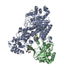
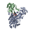
| Title | Crystal structure of Csm1-Csm4 subcomplex in the type III-A CRISPR-Csm interference complex | ||||||
 Components Components |
| ||||||
 Keywords Keywords |  IMMUNE SYSTEM / Type III-A CRISPR-Cas system / Csm1-Csm4 subcomplex IMMUNE SYSTEM / Type III-A CRISPR-Cas system / Csm1-Csm4 subcomplex | ||||||
| Function / homology |  Function and homology informationexonuclease activity / Transferases; Transferring phosphorus-containing groups; Nucleotidyltransferases / transferase activity / endonuclease activity / defense response to virus / Hydrolases; Acting on ester bonds / RNA binding / ATP binding / identical protein binding Function and homology informationexonuclease activity / Transferases; Transferring phosphorus-containing groups; Nucleotidyltransferases / transferase activity / endonuclease activity / defense response to virus / Hydrolases; Acting on ester bonds / RNA binding / ATP binding / identical protein bindingSimilarity search - Function | ||||||
| Biological species |   Thermococcus onnurineus (archaea) Thermococcus onnurineus (archaea) | ||||||
| Method | X-RAY DIFFRACTION / SYNCHROTRON / MOLECULAR REPLACEMENT / Resolution: 2.91 Å | ||||||
 Authors Authors | Jia, N. / Patel, D.J. | ||||||
 Citation Citation | Journal: Mol Cell / Year: 2019 Title: Type III-A CRISPR-Cas Csm Complexes: Assembly, Periodic RNA Cleavage, DNase Activity Regulation, and Autoimmunity. Authors: Ning Jia / Charlie Y Mo / Chongyuan Wang / Edward T Eng / Luciano A Marraffini / Dinshaw J Patel /   Abstract: Type ΙΙΙ CRISPR-Cas systems provide robust immunity against foreign RNA and DNA by sequence-specific RNase and target RNA-activated sequence-nonspecific DNase and RNase activities. We report on ...Type ΙΙΙ CRISPR-Cas systems provide robust immunity against foreign RNA and DNA by sequence-specific RNase and target RNA-activated sequence-nonspecific DNase and RNase activities. We report on cryo-EM structures of Thermococcus onnurineus Csm binary, Csm-target RNA and Csm-target RNA ternary complexes in the 3.1 Å range. The topological features of the crRNA 5'-repeat tag explains the 5'-ruler mechanism for defining target cleavage sites, with accessibility of positions -2 to -5 within the 5'-repeat serving as sensors for avoidance of autoimmunity. The Csm3 thumb elements introduce periodic kinks in the crRNA-target RNA duplex, facilitating cleavage of the target RNA with 6-nt periodicity. Key Glu residues within a Csm1 loop segment of Csm adopt a proposed autoinhibitory conformation suggestive of DNase activity regulation. These structural findings, complemented by mutational studies of key intermolecular contacts, provide insights into Csm complex assembly, mechanisms underlying RNA targeting and site-specific periodic cleavage, regulation of DNase cleavage activity, and autoimmunity suppression. | ||||||
| History |
|
- Structure visualization
| Structure viewer | Molecule: MolmilJmol/JSmol |
|---|
- Downloads & links
-Download
| PDBx/mmCIF format | 6mua.cif.gz | 204.6 KB | Display | PDBx/mmCIF format |
|---|---|---|---|---|
| PDB format | pdb6mua.ent.gz | 159.7 KB | Display | PDB format |
| PDBx/mmJSON format | 6mua.json.gz | Tree view | PDBx/mmJSON format | |
| Others |  Other downloads Other downloads |
-Validation report
| Arichive directory | https://data.pdbj.org/pub/pdb/validation_reports/mu/6muaftp://data.pdbj.org/pub/pdb/validation_reports/mu/6mua | HTTPS FTP |
|---|
-Related structure data
| Related structure data | 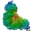 9253C 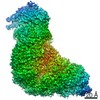 9254C 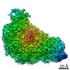 9255C  9256C  6murSC  6musC  6mutC  6muuC S: Starting model for refinement C: citing same article ( |
|---|---|
| Similar structure data |


-Links
 PDBj
PDBj- Assembly
| Deposited unit | 
| ||||||||
|---|---|---|---|---|---|---|---|---|---|
| 1 |
| ||||||||
| Unit cell |
|
-Components
| #1: Protein | Mass: 89750.602 Da / Num. of mol.: 1 Source method: isolated from a genetically manipulated source Source: (gene. exp.) Thermococcus onnurineus (archaea) / Gene: TON_0893 / Production host:  Escherichia coli (E. coli) / References: UniProt: B6YWB8 Escherichia coli (E. coli) / References: UniProt: B6YWB8 |
|---|---|
| #2: Protein | Mass: 32345.061 Da / Num. of mol.: 1 Source method: isolated from a genetically manipulated source Source: (gene. exp.) Thermococcus onnurineus (archaea) / Gene: TON_0896 / Production host: Escherichia coli (E. coli) / References: UniProt: B6YWC1 |
-Experimental details
-Experiment
| Experiment | Method: X-RAY DIFFRACTION / Number of used crystals: 1 |
|---|
- Sample preparation
| Crystal | Density Matthews: 3.06 Å3/Da / Density % sol: 59.81 % |
|---|---|
| Crystal grow | Temperature: 293 K / Method: vapor diffusion, hanging drop Details: 0.1 M phosphate-citrate pH 4.2, 5% PEG3000, 25% 1,2-propanediol, and 10% glycerol |
-Data collection
| Diffraction | Mean temperature: 100 K / Serial crystal experiment: N |
|---|---|
| Diffraction source | Source: SYNCHROTRON / Site: APS / Beamline: 24-ID-C / Wavelength: 1.2051 Å |
| Detector | Type: DECTRIS PILATUS 6M / Detector: PIXEL / Date: Jun 11, 2018 |
| Radiation | Protocol: SINGLE WAVELENGTH / Monochromatic (M) / Laue (L): M / Scattering type: x-ray |
| Radiation wavelength | Wavelength: 1.2051 Å / Relative weight: 1 |
| Reflection | Resolution: 2.9→50 Å / Num. obs: 30293 / % possible obs: 100 % / Redundancy: 16.2 % / Rpim(I) all: 0.034 / Net I/σ(I): 21.5 |
| Reflection shell | Resolution: 2.9→3 Å / Num. unique obs: 2973 / Rpim(I) all: 0.376 |
- Processing
| Software |
| ||||||||||||||||||||||||||||||||||||||||||||||||||||||||||||||||||||||||||||||||||||||||||||||||||||||||||||||||||||||||||||||||||||||||||||||||||||||||||||||||||||||||||||||||||||||
|---|---|---|---|---|---|---|---|---|---|---|---|---|---|---|---|---|---|---|---|---|---|---|---|---|---|---|---|---|---|---|---|---|---|---|---|---|---|---|---|---|---|---|---|---|---|---|---|---|---|---|---|---|---|---|---|---|---|---|---|---|---|---|---|---|---|---|---|---|---|---|---|---|---|---|---|---|---|---|---|---|---|---|---|---|---|---|---|---|---|---|---|---|---|---|---|---|---|---|---|---|---|---|---|---|---|---|---|---|---|---|---|---|---|---|---|---|---|---|---|---|---|---|---|---|---|---|---|---|---|---|---|---|---|---|---|---|---|---|---|---|---|---|---|---|---|---|---|---|---|---|---|---|---|---|---|---|---|---|---|---|---|---|---|---|---|---|---|---|---|---|---|---|---|---|---|---|---|---|---|---|---|---|---|
| Refinement | Method to determine structure: MOLECULAR REPLACEMENT Starting model: 6MUR Resolution: 2.91→49.44 Å / Cor.coef. Fo:Fc: 0.935 / Cor.coef. Fo:Fc free: 0.914 / SU B: 20.502 / SU ML: 0.38 / Cross valid method: THROUGHOUT / Details: HYDROGENS HAVE BEEN ADDED IN THE RIDING POSITIONS
| ||||||||||||||||||||||||||||||||||||||||||||||||||||||||||||||||||||||||||||||||||||||||||||||||||||||||||||||||||||||||||||||||||||||||||||||||||||||||||||||||||||||||||||||||||||||
| Solvent computation | Ion probe radii: 0.8 Å / Shrinkage radii: 0.8 Å / VDW probe radii: 1.2 Å | ||||||||||||||||||||||||||||||||||||||||||||||||||||||||||||||||||||||||||||||||||||||||||||||||||||||||||||||||||||||||||||||||||||||||||||||||||||||||||||||||||||||||||||||||||||||
| Displacement parameters | Biso mean: 91.563 Å2
| ||||||||||||||||||||||||||||||||||||||||||||||||||||||||||||||||||||||||||||||||||||||||||||||||||||||||||||||||||||||||||||||||||||||||||||||||||||||||||||||||||||||||||||||||||||||
| Refinement step | Cycle: 1 / Resolution: 2.91→49.44 Å
| ||||||||||||||||||||||||||||||||||||||||||||||||||||||||||||||||||||||||||||||||||||||||||||||||||||||||||||||||||||||||||||||||||||||||||||||||||||||||||||||||||||||||||||||||||||||
| Refine LS restraints |
|