 Movie
Movie Controller

[English] 日本語
 Yorodumi
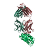
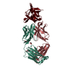
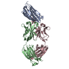
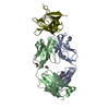
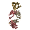
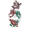
- PDB-6mto: Crystal structure of VRC42.01 Fab in complex with T117-F MPER scaffold -
+ Open data

- Basic information
| Entry | Database: PDB / ID: 6mto | ||||||
|---|---|---|---|---|---|---|---|
| Title | Crystal structure of VRC42.01 Fab in complex with T117-F MPER scaffold | ||||||
 Components Components |
| ||||||
 Keywords Keywords |  IMMUNE SYSTEM / Anti-HIV-1 human antibody / MPER / gp41 IMMUNE SYSTEM / Anti-HIV-1 human antibody / MPER / gp41 | ||||||
| Function / homology | Mannitol-specific EII; Chain A / Mannitol-specific EII; Chain A / Immunoglobulins / Immunoglobulin-like / Sandwich / 3-Layer(aba) Sandwich / Mainly Beta / Alpha Beta Function and homology information Function and homology information | ||||||
| Biological species |  Homo sapiens (human) Homo sapiens (human)  Human immunodeficiency virus 1 Human immunodeficiency virus 1 | ||||||
| Method | X-RAY DIFFRACTION / SYNCHROTRON / MOLECULAR REPLACEMENT / Resolution: 2.634 Å | ||||||
 Authors Authors | Kwon, Y.D. / Druz, A. / Law, W.H. / Peng, D. / Zhang, B. / Doria-Rose, N.A. / Kwong, P.D. | ||||||
| Funding support |  United States, 1items United States, 1items
| ||||||
 Citation Citation | Journal: Immunity / Year: 2019 Title: Longitudinal Analysis Reveals Early Development of Three MPER-Directed Neutralizing Antibody Lineages from an HIV-1-Infected Individual. Authors: Krebs, S.J. / Kwon, Y.D. / Schramm, C.A. / Law, W.H. / Donofrio, G. / Zhou, K.H. / Gift, S. / Dussupt, V. / Georgiev, I.S. / Schatzle, S. / McDaniel, J.R. / Lai, Y.T. / Sastry, M. / Zhang, B. ...Authors: Krebs, S.J. / Kwon, Y.D. / Schramm, C.A. / Law, W.H. / Donofrio, G. / Zhou, K.H. / Gift, S. / Dussupt, V. / Georgiev, I.S. / Schatzle, S. / McDaniel, J.R. / Lai, Y.T. / Sastry, M. / Zhang, B. / Jarosinski, M.C. / Ransier, A. / Chenine, A.L. / Asokan, M. / Bailer, R.T. / Bose, M. / Cagigi, A. / Cale, E.M. / Chuang, G.Y. / Darko, S. / Driscoll, J.I. / Druz, A. / Gorman, J. / Laboune, F. / Louder, M.K. / McKee, K. / Mendez, L. / Moody, M.A. / O'Sullivan, A.M. / Owen, C. / Peng, D. / Rawi, R. / Sanders-Buell, E. / Shen, C.H. / Shiakolas, A.R. / Stephens, T. / Tsybovsky, Y. / Tucker, C. / Verardi, R. / Wang, K. / Zhou, J. / Zhou, T. / Georgiou, G. / Alam, S.M. / Haynes, B.F. / Rolland, M. / Matyas, G.R. / Polonis, V.R. / McDermott, A.B. / Douek, D.C. / Shapiro, L. / Tovanabutra, S. / Michael, N.L. / Mascola, J.R. / Robb, M.L. / Kwong, P.D. / Doria-Rose, N.A. | ||||||
| History |
|
- Structure visualization
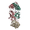
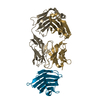
| Structure viewer | Molecule: MolmilJmol/JSmol |
|---|
- Downloads & links
-Download
| PDBx/mmCIF format | 6mto.cif.gz | 201.2 KB | Display | PDBx/mmCIF format |
|---|---|---|---|---|
| PDB format | pdb6mto.ent.gz | 162.3 KB | Display | PDB format |
| PDBx/mmJSON format | 6mto.json.gz | Tree view | PDBx/mmJSON format | |
| Others |  Other downloads Other downloads |
-Validation report
| Arichive directory | https://data.pdbj.org/pub/pdb/validation_reports/mt/6mtoftp://data.pdbj.org/pub/pdb/validation_reports/mt/6mto | HTTPS FTP |
|---|
-Related structure data
| Related structure data | 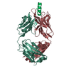 6mtpSC 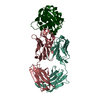 6mtqC 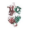 6mtrC  6mtsC  6mttC S: Starting model for refinement C: citing same article ( |
|---|---|
| Similar structure data |


-Links
 PDBj
PDBj

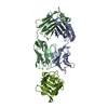
- Assembly
| Deposited unit | 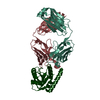
| ||||||||
|---|---|---|---|---|---|---|---|---|---|
| 1 |
| ||||||||
| Unit cell |
|
-Components
| #1: Antibody | Mass: 23482.959 Da / Num. of mol.: 1 Source method: isolated from a genetically manipulated source Source: (gene. exp.) Homo sapiens (human) / Plasmid: pVRC8400 / Cell line (production host): HEK293F / Production host: Homo sapiens (human) |
|---|---|
| #2: Antibody | Mass: 23717.805 Da / Num. of mol.: 1 Source method: isolated from a genetically manipulated source Source: (gene. exp.) Homo sapiens (human) / Plasmid: pVRC8400 / Cell line (production host): HEK293F / Production host: Homo sapiens (human) |
| #3: Protein | Mass: 18218.713 Da / Num. of mol.: 1 Source method: isolated from a genetically manipulated source Source: (gene. exp.) Human immunodeficiency virus 1 / Plasmid: pVRC8400 / Cell line (production host): HEK293F / Production host: Homo sapiens (human) |
| #4: Water | ChemComp-HOH / Water Mass: 18.015 Da / Num. of mol.: 9 / Source method: isolated from a natural source / Formula: H2O Mass: 18.015 Da / Num. of mol.: 9 / Source method: isolated from a natural source / Formula: H2O |
-Experimental details
-Experiment
| Experiment | Method: X-RAY DIFFRACTION / Number of used crystals: 1 |
|---|
- Sample preparation
| Crystal | Density Matthews: 2.48 Å3/Da / Density % sol: 50.45 % |
|---|---|
| Crystal grow | Temperature: 293 K / Method: vapor diffusion, hanging drop / pH: 5.5 / Details: 10% PEG 400, 2% PEG 3350, 0.1M Na Acetate 5.5 |
-Data collection
| Diffraction | Mean temperature: 100 K / Serial crystal experiment: N |
|---|---|
| Diffraction source | Source: SYNCHROTRON / Site: APS / Beamline: 22-ID / Wavelength: 1 Å |
| Detector | Type: MARMOSAIC 300 mm CCD / Detector: CCD / Date: Jul 6, 2017 |
| Radiation | Monochromator: double crystal - liquid nitrogen cooled / Protocol: SINGLE WAVELENGTH / Monochromatic (M) / Laue (L): M / Scattering type: x-ray |
| Radiation wavelength | Wavelength: 1 Å / Relative weight: 1 |
| Reflection | Resolution: 2.63→50 Å / Num. obs: 17700 / % possible obs: 92.4 % / Redundancy: 3.4 % / CC1/2: 0.867 / Rmerge(I) obs: 0.104 / Rpim(I) all: 0.062 / Net I/σ(I): 11.3 |
| Reflection shell | Resolution: 2.63→2.7 Å / Rmerge(I) obs: 0.556 / Num. unique all: 580 / CC1/2: 0.555 / Rpim(I) all: 0.479 |
- Processing
| Software |
| ||||||||||||||||||||||||||||||||||||||||||||||||||||||||||||||||||||||||||||||||||||||||||||||||||||||||||||||||||||||||||||||||||||||||||||||||||||||
|---|---|---|---|---|---|---|---|---|---|---|---|---|---|---|---|---|---|---|---|---|---|---|---|---|---|---|---|---|---|---|---|---|---|---|---|---|---|---|---|---|---|---|---|---|---|---|---|---|---|---|---|---|---|---|---|---|---|---|---|---|---|---|---|---|---|---|---|---|---|---|---|---|---|---|---|---|---|---|---|---|---|---|---|---|---|---|---|---|---|---|---|---|---|---|---|---|---|---|---|---|---|---|---|---|---|---|---|---|---|---|---|---|---|---|---|---|---|---|---|---|---|---|---|---|---|---|---|---|---|---|---|---|---|---|---|---|---|---|---|---|---|---|---|---|---|---|---|---|---|---|---|
| Refinement | Method to determine structure: MOLECULAR REPLACEMENT Starting model: 6MTP, VRC42.04 Resolution: 2.634→30.263 Å / SU ML: 0.41 / Cross valid method: FREE R-VALUE / σ(F): 1.35 / Phase error: 33.55
| ||||||||||||||||||||||||||||||||||||||||||||||||||||||||||||||||||||||||||||||||||||||||||||||||||||||||||||||||||||||||||||||||||||||||||||||||||||||
| Solvent computation | Shrinkage radii: 0.9 Å / VDW probe radii: 1.11 Å | ||||||||||||||||||||||||||||||||||||||||||||||||||||||||||||||||||||||||||||||||||||||||||||||||||||||||||||||||||||||||||||||||||||||||||||||||||||||
| Refinement step | Cycle: LAST / Resolution: 2.634→30.263 Å
| ||||||||||||||||||||||||||||||||||||||||||||||||||||||||||||||||||||||||||||||||||||||||||||||||||||||||||||||||||||||||||||||||||||||||||||||||||||||
| Refine LS restraints |
| ||||||||||||||||||||||||||||||||||||||||||||||||||||||||||||||||||||||||||||||||||||||||||||||||||||||||||||||||||||||||||||||||||||||||||||||||||||||
| LS refinement shell |
| ||||||||||||||||||||||||||||||||||||||||||||||||||||||||||||||||||||||||||||||||||||||||||||||||||||||||||||||||||||||||||||||||||||||||||||||||||||||
| Refinement TLS params. | Method: refined / Refine-ID: X-RAY DIFFRACTION
| ||||||||||||||||||||||||||||||||||||||||||||||||||||||||||||||||||||||||||||||||||||||||||||||||||||||||||||||||||||||||||||||||||||||||||||||||||||||
| Refinement TLS group |
|
