 Movie
Movie Controller


+ Open data

- Basic information
| Entry | Database: PDB / ID: 6mrw | ||||||
|---|---|---|---|---|---|---|---|
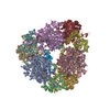
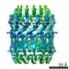
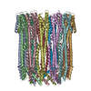
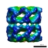
| Title | 14-meric ClyA pore complex | ||||||
 Components Components | Hemolysin E, chromosomal | ||||||
 Keywords Keywords |  MEMBRANE PROTEIN / Toxin / Pore-forming toxin MEMBRANE PROTEIN / Toxin / Pore-forming toxin | ||||||
| Function / homology |  Function and homology information Function and homology informationmodulation of apoptotic process in another organism / hemolysis in another organism / toxin activity / periplasmic space / host cell plasma membrane / extracellular region / membrane / identical protein bindingSimilarity search - Function | ||||||
| Biological species |  Escherichia coli (E. coli) Escherichia coli (E. coli) | ||||||
| Method | ELECTRON MICROSCOPY / single particle reconstruction / cryo EM / Resolution: 4.3 Å | ||||||
 Authors Authors | Peng, W. / de Souza Santos, M. / Li, Y. / Tomchick, D.R. / Orth, K. | ||||||
 Citation Citation | Journal: PLoS One / Year: 2019 Title: High-resolution cryo-EM structures of the E. coli hemolysin ClyA oligomers. Authors: Wei Peng / Marcela de Souza Santos / Yang Li / Diana R Tomchick / Kim Orth /  Abstract: Pore-forming proteins (PFPs) represent a functionally important protein family, that are found in organisms from viruses to humans. As a major branch of PFPs, bacteria pore-forming toxins (PFTs) ...Pore-forming proteins (PFPs) represent a functionally important protein family, that are found in organisms from viruses to humans. As a major branch of PFPs, bacteria pore-forming toxins (PFTs) permeabilize membranes and usually cause the death of target cells. E. coli hemolysin ClyA is the first member with the pore complex structure solved among α-PFTs, employing α-helices as transmembrane elements. ClyA is proposed to form pores composed of various numbers of protomers. With high-resolution cryo-EM structures, we observe that ClyA pore complexes can exist as newly confirmed oligomers of a tridecamer and a tetradecamer, at estimated resolutions of 3.2 Å and 4.3 Å, respectively. The 2.8 Å cryo-EM structure of a dodecamer dramatically improves the existing structural model. Structural analysis indicates that protomers from distinct oligomers resemble each other and neighboring protomers adopt a conserved interaction mode. We also show a stabilized intermediate state of ClyA during the transition process from soluble monomers to pore complexes. Unexpectedly, even without the formation of mature pore complexes, ClyA can permeabilize membranes and allow leakage of particles less than ~400 Daltons. In addition, we are the first to show that ClyA forms pore complexes in the presence of cholesterol within artificial liposomes. These findings provide new mechanistic insights into the dynamic process of pore assembly for the prototypical α-PFT ClyA. | ||||||
| History |
|
- Structure visualization
| Movie |
Movie viewer |
|---|---|
| Structure viewer | Molecule: MolmilJmol/JSmol |
- Downloads & links
-Download
| PDBx/mmCIF format | 6mrw.cif.gz | 915.5 KB | Display | PDBx/mmCIF format |
|---|---|---|---|---|
| PDB format | pdb6mrw.ent.gz | 724.7 KB | Display | PDB format |
| PDBx/mmJSON format | 6mrw.json.gz | Tree view | PDBx/mmJSON format | |
| Others |  Other downloads Other downloads |
-Validation report
| Arichive directory | https://data.pdbj.org/pub/pdb/validation_reports/mr/6mrwftp://data.pdbj.org/pub/pdb/validation_reports/mr/6mrw | HTTPS FTP |
|---|
-Related structure data
| Related structure data |  9214MC  9212C  9213C  6mrtC  6mruC M: map data used to model this data C: citing same article ( |
|---|---|
| Similar structure data |

-Links
 PDBj
PDBj- Assembly
| Deposited unit | 
|
|---|---|
| 1 |
|
-Components
| #1: Protein | Mass: 36131.816 Da / Num. of mol.: 14 Source method: isolated from a genetically manipulated source Source: (gene. exp.) Escherichia coli (strain K12) (bacteria)Strain: K12 / Gene: hlyE, clyA, hpr, sheA, ycgD, b1182, JW5181 / Production host: Escherichia coli (E. coli) / References: UniProt: P77335 |
|---|
-Experimental details
-Experiment
| Experiment | Method: ELECTRON MICROSCOPY |
|---|---|
| EM experiment | Aggregation state: PARTICLE / 3D reconstruction method: single particle reconstruction |
- Sample preparation
| Component | Name: 14-meric ClyA pore complex / Type: COMPLEX / Entity ID: all / Source: RECOMBINANT |
|---|---|
| Molecular weight | Value: 0.48 MDa / Experimental value: YES |
| Source (natural) | Organism: Escherichia coli (E. coli) |
| Source (recombinant) | Organism: Escherichia coli (E. coli) |
| Buffer solution | pH: 8 |
| Specimen | Embedding applied: NO / Shadowing applied: NO / Staining applied: NO / Vitrification applied: YES |
| Specimen support | Details: unspecified |
| Vitrification | Cryogen name: ETHANE |
- Electron microscopy imaging
| Experimental equipment |  Model: Titan Krios / Image courtesy: FEI Company |
|---|---|
| Microscopy | Model: FEI TITAN KRIOS |
| Electron gun | Electron source: FIELD EMISSION GUN / Accelerating voltage: 300 kV / Illumination mode: FLOOD BEAM |
| Electron lens | Mode: BRIGHT FIELDBright-field microscopy |
| Image recording | Electron dose: 50 e/Å2 / Detector mode: SUPER-RESOLUTION / Film or detector model: GATAN K2 SUMMIT (4k x 4k) |
- Processing
| CTF correction | Type: NONE |
|---|---|
| 3D reconstruction | Resolution: 4.3 Å / Resolution method: FSC 0.143 CUT-OFF / Num. of particles: 30904 / Symmetry type: POINT |
