 Movie
Movie Controller

[English] 日本語
 Yorodumi
- PDB-6mql: Crystal Structure of GTPase Domain of Human Septin 12 Mutant T89M -
+ Open data

- Basic information
| Entry | Database: PDB / ID: 6mql | |||||||||
|---|---|---|---|---|---|---|---|---|---|---|
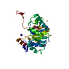
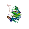
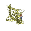
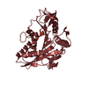
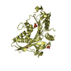
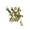
| Title | Crystal Structure of GTPase Domain of Human Septin 12 Mutant T89M | |||||||||
 Components Components | Septin-12 | |||||||||
 Keywords Keywords | STRUCTURAL PROTEIN / cytoskeleton component septin GTPase spermatogenesis | |||||||||
| Function / homology |  Function and homology information Function and homology informationsperm annulus / septin complex / cytoskeleton-dependent cytokinesis / septin ring / cell division site / cleavage furrow / stress fiber / phosphatidylinositol binding / spindle / GDP binding ...sperm annulus / septin complex / cytoskeleton-dependent cytokinesis / septin ring / cell division site / cleavage furrow / stress fiber / phosphatidylinositol binding / spindle / GDP binding / microtubule cytoskeleton / midbody / spermatogenesis / molecular adaptor activity / cell differentiation / GTPase activity / GTP binding / perinuclear region of cytoplasm / protein homodimerization activity / identical protein binding / nucleusSimilarity search - Function | |||||||||
| Biological species |  Homo sapiens (human) Homo sapiens (human) | |||||||||
| Method | X-RAY DIFFRACTION / SYNCHROTRON / MOLECULAR REPLACEMENT / Resolution: 2.17 Å | |||||||||
 Authors Authors | Castro, D.K.S.V. / Pereira, H.M. / Brandao-Neto, J. / Ulian, A.P.U. / Garratt, R.C. | |||||||||
| Funding support |  Brazil, 2items Brazil, 2items
| |||||||||
 Citation Citation | Journal: To Be Published Title: Crystal Structure of GTPase Domain of Human Septin 12 Authors: Castro, D.K.S.V. / Pereira, H.M. / Brandao-Neto, J. / Ulian, A.P.U. / Garratt, R.C. | |||||||||
| History |
|
- Structure visualization
| Structure viewer | Molecule: MolmilJmol/JSmol |
|---|
- Downloads & links
-Download
| PDBx/mmCIF format | 6mql.cif.gz | 125.8 KB | Display | PDBx/mmCIF format |
|---|---|---|---|---|
| PDB format | pdb6mql.ent.gz | 95.3 KB | Display | PDB format |
| PDBx/mmJSON format | 6mql.json.gz | Tree view | PDBx/mmJSON format | |
| Others |  Other downloads Other downloads |
-Validation report
| Arichive directory | https://data.pdbj.org/pub/pdb/validation_reports/mq/6mqlftp://data.pdbj.org/pub/pdb/validation_reports/mq/6mql | HTTPS FTP |
|---|
-Related structure data
| Related structure data |  6mq9S S: Starting model for refinement |
|---|---|
| Similar structure data |


-Links
 PDBj
PDBj- Assembly
| Deposited unit | 
| ||||||||
|---|---|---|---|---|---|---|---|---|---|
| 1 |
| ||||||||
| 2 | 
| ||||||||
| Unit cell |
|
-Components
| #1: Protein | Mass: 34126.195 Da / Num. of mol.: 1 / Mutation: T89M Source method: isolated from a genetically manipulated source Source: (gene. exp.) Homo sapiens (human) / Gene: SEPT12 / Production host:  Escherichia coli (E. coli) / Strain (production host): Rosetta DE3 / References: UniProt: Q8IYM1 Escherichia coli (E. coli) / Strain (production host): Rosetta DE3 / References: UniProt: Q8IYM1 |
|---|---|
| #2: Chemical | ChemComp-GDP / Guanosine diphosphate  Type: RNA linking / Mass: 443.201 Da / Num. of mol.: 1 / Source method: obtained synthetically / Formula: C10H15N5O11P2 / Comment: GDP, energy-carrying molecule*YM Type: RNA linking / Mass: 443.201 Da / Num. of mol.: 1 / Source method: obtained synthetically / Formula: C10H15N5O11P2 / Comment: GDP, energy-carrying molecule*YM |
| #3: Chemical | ChemComp-MG /   Mass: 24.305 Da / Num. of mol.: 1 / Source method: obtained synthetically / Formula: Mg Mass: 24.305 Da / Num. of mol.: 1 / Source method: obtained synthetically / Formula: Mg |
| #4: Water | ChemComp-HOH / Water Mass: 18.015 Da / Num. of mol.: 45 / Source method: isolated from a natural source / Formula: H2O Mass: 18.015 Da / Num. of mol.: 45 / Source method: isolated from a natural source / Formula: H2O |
-Experimental details
-Experiment
| Experiment | Method: X-RAY DIFFRACTION / Number of used crystals: 1 |
|---|
- Sample preparation
| Crystal | Density Matthews: 2.21 Å3/Da / Density % sol: 44.47 % |
|---|---|
| Crystal grow | Temperature: 291 K / Method: vapor diffusion, sitting drop / pH: 8.5 Details: 10% w/v PEG 4000, 20% v/v glycerol, 0.1 M bicine/Trizma base pH 8.5, 30mM of each: diethyleneglycol, triethyleneglycol, tetraethyleneglycol, pentaethyleneglycol |
-Data collection
| Diffraction | Mean temperature: 100 K / Serial crystal experiment: N |
|---|---|
| Diffraction source | Source: SYNCHROTRON / Site: Diamond  / Beamline: I24 / Wavelength: 0.9686 Å / Beamline: I24 / Wavelength: 0.9686 Å |
| Detector | Type: DECTRIS PILATUS3 6M / Detector: PIXEL / Date: Oct 15, 2017 |
| Radiation | Protocol: SINGLE WAVELENGTH / Monochromatic (M) / Laue (L): M / Scattering type: x-ray |
| Radiation wavelength | Wavelength: 0.9686 Å / Relative weight: 1 |
| Reflection | Resolution: 2.17→28.25 Å / Num. obs: 15878 / % possible obs: 99.6 % / Redundancy: 6.5 % / CC1/2: 0.995 / Rpim(I) all: 0.059 / Net I/σ(I): 8.7 |
| Reflection shell | Resolution: 2.17→2.23 Å / Redundancy: 6.7 % / Mean I/σ(I) obs: 1.7 / Num. unique obs: 1161 / CC1/2: 0.528 / Rpim(I) all: 0.488 / % possible all: 99.6 |
- Processing
| Software |
| |||||||||||||||||||||||||||||||||||||||||||||||||||||||||||||||||||||||||||||||||||||||||||||||||||||||||||||||||||||||||||||||||||||||||||||||||||||||||||||||||||||||||||||||
|---|---|---|---|---|---|---|---|---|---|---|---|---|---|---|---|---|---|---|---|---|---|---|---|---|---|---|---|---|---|---|---|---|---|---|---|---|---|---|---|---|---|---|---|---|---|---|---|---|---|---|---|---|---|---|---|---|---|---|---|---|---|---|---|---|---|---|---|---|---|---|---|---|---|---|---|---|---|---|---|---|---|---|---|---|---|---|---|---|---|---|---|---|---|---|---|---|---|---|---|---|---|---|---|---|---|---|---|---|---|---|---|---|---|---|---|---|---|---|---|---|---|---|---|---|---|---|---|---|---|---|---|---|---|---|---|---|---|---|---|---|---|---|---|---|---|---|---|---|---|---|---|---|---|---|---|---|---|---|---|---|---|---|---|---|---|---|---|---|---|---|---|---|---|---|---|---|
| Refinement | Method to determine structure: MOLECULAR REPLACEMENT Starting model: 6MQ9 Resolution: 2.17→28.245 Å / SU ML: 0.37 / Cross valid method: THROUGHOUT / σ(F): 1.35 / Phase error: 30.64
| |||||||||||||||||||||||||||||||||||||||||||||||||||||||||||||||||||||||||||||||||||||||||||||||||||||||||||||||||||||||||||||||||||||||||||||||||||||||||||||||||||||||||||||||
| Solvent computation | Shrinkage radii: 0.9 Å / VDW probe radii: 1.11 Å | |||||||||||||||||||||||||||||||||||||||||||||||||||||||||||||||||||||||||||||||||||||||||||||||||||||||||||||||||||||||||||||||||||||||||||||||||||||||||||||||||||||||||||||||
| Displacement parameters | Biso max: 130.11 Å2 / Biso mean: 55.9275 Å2 / Biso min: 28.67 Å2 | |||||||||||||||||||||||||||||||||||||||||||||||||||||||||||||||||||||||||||||||||||||||||||||||||||||||||||||||||||||||||||||||||||||||||||||||||||||||||||||||||||||||||||||||
| Refinement step | Cycle: final / Resolution: 2.17→28.245 Å
| |||||||||||||||||||||||||||||||||||||||||||||||||||||||||||||||||||||||||||||||||||||||||||||||||||||||||||||||||||||||||||||||||||||||||||||||||||||||||||||||||||||||||||||||
| Refine LS restraints |
| |||||||||||||||||||||||||||||||||||||||||||||||||||||||||||||||||||||||||||||||||||||||||||||||||||||||||||||||||||||||||||||||||||||||||||||||||||||||||||||||||||||||||||||||
| LS refinement shell | Refine-ID: X-RAY DIFFRACTION / Rfactor Rfree error: 0 / Total num. of bins used: 5 / % reflection obs: 100 %
| |||||||||||||||||||||||||||||||||||||||||||||||||||||||||||||||||||||||||||||||||||||||||||||||||||||||||||||||||||||||||||||||||||||||||||||||||||||||||||||||||||||||||||||||
| Refinement TLS params. | Method: refined / Refine-ID: X-RAY DIFFRACTION
| |||||||||||||||||||||||||||||||||||||||||||||||||||||||||||||||||||||||||||||||||||||||||||||||||||||||||||||||||||||||||||||||||||||||||||||||||||||||||||||||||||||||||||||||
| Refinement TLS group |
|
