Mass: 18.015 Da / Num. of mol.: 1063 / Source method: isolated from a natural source / Formula: H2O
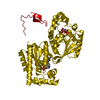
Has ligand of interest
N
Sequence details
THE SEQUENCE OF THIS PROTEIN WAS NOT AVAILABLE AT THE UNIPROT KNOWLEDGEBASE DATABASE (UNIPROTKB) AT ...THE SEQUENCE OF THIS PROTEIN WAS NOT AVAILABLE AT THE UNIPROT KNOWLEDGEBASE DATABASE (UNIPROTKB) AT THE TIME OF DEPOSITION. AUTHORS STATE THAT THE GENEBANK ACCESSION NUMBER IS EOK08638.1. FOR THE PROTEIN.
-
Experimental details
-
Experiment
Experiment
Method: X-RAY DIFFRACTION / Number of used crystals: 1
-
Sample preparation
Crystal
Density Matthews: 2.5 Å3/Da / Density % sol: 50.87 %
Crystal grow
Temperature: 293 K / Method: vapor diffusion, hanging drop / Details: 25% PEG3350, 0.2 M ammonium citrate
-
Data collection
Diffraction
Mean temperature: 100 K / Serial crystal experiment: N
Type: DECTRIS PILATUS3 S 6M / Detector: PIXEL / Date: Nov 24, 2019
Radiation
Protocol: SINGLE WAVELENGTH / Monochromatic (M) / Laue (L): M / Scattering type: x-ray
Radiation wavelength
Wavelength: 1 Å / Relative weight: 1
Reflection
Resolution: 1.4→50 Å / Num. obs: 201712 / % possible obs: 100 % / Redundancy: 12.7 % / CC1/2: 1 / Rmerge(I) obs: 0.068 / Net I/σ(I): 18.3
Reflection shell
Resolution: 1.4→1.48 Å / Redundancy: 13.1 % / Rmerge(I) obs: 0.877 / Mean I/σ(I) obs: 2.7 / Num. unique obs: 29211 / CC1/2: 0.884 / % possible all: 100
-
Processing
Software
Name
Version
Classification
REFMAC
5.8.0258
refinement
XDS
datareduction
XDS
datascaling
PHENIX
phasing
Refinement
Method to determine structure: SAD / Resolution: 1.4→46.123 Å / Cor.coef. Fo:Fc: 0.973 / Cor.coef. Fo:Fc free: 0.965 / SU B: 1.103 / SU ML: 0.043 / Cross valid method: FREE R-VALUE / ESU R: 0.056 / ESU R Free: 0.058 Details: Hydrogens have been added in their riding positions
Rfactor
Num. reflection
% reflection
Rfree
0.1951
9974
4.963 %
Rwork
0.1715
-
-
all
0.173
-
-
obs
-
200987
99.67 %
Solvent computation
Ion probe radii: 0.8 Å / Shrinkage radii: 0.8 Å / VDW probe radii: 1.2 Å
Displacement parameters
Biso mean: 22.259 Å2
Baniso -1
Baniso -2
Baniso -3
1-
0.093 Å2
0 Å2
0 Å2
2-
-
-0.02 Å2
0 Å2
3-
-
-
-0.073 Å2
Refinement step
Cycle: LAST / Resolution: 1.4→46.123 Å
Protein
Nucleic acid
Ligand
Solvent
Total
Num. atoms
7216
0
36
1063
8315
Refine LS restraints
Refine-ID
Type
Dev ideal
Dev ideal target
Number
X-RAY DIFFRACTION
r_bond_refined_d
0.008
0.013
7518
X-RAY DIFFRACTION
r_bond_other_d
0.001
0.017
6639
X-RAY DIFFRACTION
r_angle_refined_deg
1.47
1.65
10205
X-RAY DIFFRACTION
r_angle_other_deg
1.408
1.578
15521
X-RAY DIFFRACTION
r_dihedral_angle_1_deg
7.335
5
946
X-RAY DIFFRACTION
r_dihedral_angle_2_deg
34.411
24.845
386
X-RAY DIFFRACTION
r_dihedral_angle_3_deg
12.078
15
1223
X-RAY DIFFRACTION
r_dihedral_angle_4_deg
17.901
15
21
X-RAY DIFFRACTION
r_chiral_restr
0.073
0.2
965
X-RAY DIFFRACTION
r_gen_planes_refined
0.008
0.02
8593
X-RAY DIFFRACTION
r_gen_planes_other
0.001
0.02
1526
X-RAY DIFFRACTION
r_nbd_refined
0.207
0.2
1401
X-RAY DIFFRACTION
r_symmetry_nbd_other
0.179
0.2
6245
X-RAY DIFFRACTION
r_nbtor_refined
0.169
0.2
3654
X-RAY DIFFRACTION
r_symmetry_nbtor_other
0.076
0.2
3188
X-RAY DIFFRACTION
r_xyhbond_nbd_refined
0.134
0.2
759
X-RAY DIFFRACTION
r_symmetry_xyhbond_nbd_other
0.119
0.2
2
X-RAY DIFFRACTION
r_symmetry_nbd_refined
0.26
0.2
17
X-RAY DIFFRACTION
r_nbd_other
0.241
0.2
43
X-RAY DIFFRACTION
r_symmetry_xyhbond_nbd_refined
0.169
0.2
38
X-RAY DIFFRACTION
r_mcbond_it
1.391
2.218
3751
X-RAY DIFFRACTION
r_mcbond_other
1.39
2.217
3750
X-RAY DIFFRACTION
r_mcangle_it
2.108
3.325
4708
X-RAY DIFFRACTION
r_mcangle_other
2.108
3.326
4709
X-RAY DIFFRACTION
r_scbond_it
2.041
2.395
3767
X-RAY DIFFRACTION
r_scbond_other
2.041
2.396
3768
X-RAY DIFFRACTION
r_scangle_it
3.091
3.511
5497
X-RAY DIFFRACTION
r_scangle_other
3.091
3.511
5498
X-RAY DIFFRACTION
r_lrange_it
4.693
26.895
8739
X-RAY DIFFRACTION
r_lrange_other
4.44
26.101
8452
LS refinement shell
Resolution (Å)
Rfactor Rfree
Num. reflection Rfree
Rfactor Rwork
Num. reflection Rwork
Refine-ID
% reflection obs (%)
1.4-1.436
0.286
663
0.263
14118
X-RAY DIFFRACTION
99.9729
1.436-1.476
0.303
685
0.294
13695
X-RAY DIFFRACTION
99.7434
1.476-1.518
0.271
712
0.258
13237
X-RAY DIFFRACTION
99.6357
1.518-1.565
0.298
640
0.271
12911
X-RAY DIFFRACTION
99.6104
1.565-1.616
0.222
667
0.202
12546
X-RAY DIFFRACTION
99.8791
1.616-1.673
0.215
642
0.19
12119
X-RAY DIFFRACTION
99.8045
1.673-1.736
0.198
627
0.182
11688
X-RAY DIFFRACTION
99.7812
1.736-1.807
0.21
599
0.187
11248
X-RAY DIFFRACTION
99.6887
1.807-1.887
0.222
548
0.183
10871
X-RAY DIFFRACTION
99.6683
1.887-1.98
0.222
515
0.2
10171
X-RAY DIFFRACTION
97.893
1.98-2.087
0.209
557
0.177
9816
X-RAY DIFFRACTION
99.4917
2.087-2.213
0.189
436
0.161
9385
X-RAY DIFFRACTION
99.8983
2.213-2.366
0.202
501
0.166
8744
X-RAY DIFFRACTION
99.4086
2.366-2.555
0.177
482
0.15
8162
X-RAY DIFFRACTION
99.9191
2.555-2.799
0.178
415
0.149
7592
X-RAY DIFFRACTION
99.9251
2.799-3.128
0.182
344
0.151
6901
X-RAY DIFFRACTION
99.9448
3.128-3.611
0.18
304
0.152
6146
X-RAY DIFFRACTION
100
3.611-4.42
0.161
256
0.141
5226
X-RAY DIFFRACTION
99.9635
4.42-6.24
0.145
241
0.145
4068
X-RAY DIFFRACTION
100
6.24-46.123
0.187
140
0.167
2369
X-RAY DIFFRACTION
99.6426
+
About Yorodumi
-
News
-
Feb 9, 2022. New format data for meta-information of EMDB entries
New format data for meta-information of EMDB entries
Version 3 of the EMDB header file is now the official format.
The previous official version 1.9 will be removed from the archive.
In the structure databanks used in Yorodumi, some data are registered as the other names, "COVID-19 virus" and "2019-nCoV". Here are the details of the virus and the list of structure data.
Jan 31, 2019. EMDB accession codes are about to change! (news from PDBe EMDB page)
EMDB accession codes are about to change! (news from PDBe EMDB page)
The allocation of 4 digits for EMDB accession codes will soon come to an end. Whilst these codes will remain in use, new EMDB accession codes will include an additional digit and will expand incrementally as the available range of codes is exhausted. The current 4-digit format prefixed with “EMD-” (i.e. EMD-XXXX) will advance to a 5-digit format (i.e. EMD-XXXXX), and so on. It is currently estimated that the 4-digit codes will be depleted around Spring 2019, at which point the 5-digit format will come into force.
The EM Navigator/Yorodumi systems omit the EMD- prefix.
Related info.:Q: What is EMD? / ID/Accession-code notation in Yorodumi/EM Navigator
Yorodumi is a browser for structure data from EMDB, PDB, SASBDB, etc.
This page is also the successor to EM Navigator detail page, and also detail information page/front-end page for Omokage search.
The word "yorodu" (or yorozu) is an old Japanese word meaning "ten thousand". "mi" (miru) is to see.
Related info.:EMDB / PDB / SASBDB / Comparison of 3 databanks / Yorodumi Search / Aug 31, 2016. New EM Navigator & Yorodumi / Yorodumi Papers / Jmol/JSmol / Function and homology information / Changes in new EM Navigator and Yorodumi
 Movie
Movie Controller


 Open data
 Basic information
 Components
 Keywords
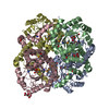
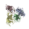
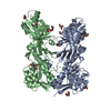
 HYDROLASE /
  Function and homology information
 Authors
 Japan, 1items
  Citation
 Structure visualization
 Downloads & links
 Other downloads
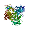


 PDBj
PDBj Assembly

 Mass: 62.068 Da / Num. of mol.: 9 / Source method: obtained synthetically / Formula: C2H6O2
 Mass: 18.015 Da / Num. of mol.: 1063 / Source method: isolated from a natural source / Formula: H2O
 Sample preparation
 Processing