 Movie
Movie Controller

[English] 日本語
 Yorodumi
- PDB-6m3y: Crystal structure of pilus adhesin, SpaC from Lactobacillus rhamn... -
+ Open data

- Basic information
| Entry | Database: PDB / ID: 6m3y | ||||||
|---|---|---|---|---|---|---|---|
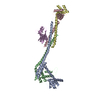
| Title | Crystal structure of pilus adhesin, SpaC from Lactobacillus rhamnosus GG - open conformation | ||||||
 Components Components | Pilus assembly protein | ||||||
 Keywords Keywords |  CELL ADHESION / Pilus adhesin / Tip pilin / vWFA domain / SpaCBA pilus / sortase / Lactobacillus rhamnosus GG / Pili / Fimbria / Probiotics / SpaC / lectin / surface protein. CELL ADHESION / Pilus adhesin / Tip pilin / vWFA domain / SpaCBA pilus / sortase / Lactobacillus rhamnosus GG / Pili / Fimbria / Probiotics / SpaC / lectin / surface protein. | ||||||
| Function / homology | Prealbumin-like fold domain / Prealbumin-like fold domain / von Willebrand factor type A domain / von Willebrand factor (vWF) type A domain / VWFA domain profile. / von Willebrand factor, type A / von Willebrand factor A-like domain superfamily / Immunoglobulin-like fold / Pilus assembly protein Function and homology information Function and homology information | ||||||
| Biological species |  Lactobacillus rhamnosus (bacteria) Lactobacillus rhamnosus (bacteria) | ||||||
| Method | X-RAY DIFFRACTION / SYNCHROTRON / MOLECULAR REPLACEMENT / Resolution: 1.93 Å | ||||||
 Authors Authors | Kant, A. / Palva, A. / von Ossowski, I. / Krishnan, V. | ||||||
| Funding support |  India, 1items India, 1items
| ||||||
 Citation Citation | Journal: J.Struct.Biol. / Year: 2020 Title: Crystal structure of lactobacillar SpaC reveals an atypical five-domain pilus tip adhesin: Exposing its substrate-binding and assembly in SpaCBA pili. Authors: Kant, A. / Palva, A. / von Ossowski, I. / Krishnan, V. | ||||||
| History |
|
- Structure visualization
| Structure viewer | Molecule: MolmilJmol/JSmol |
|---|
- Downloads & links
-Download
| PDBx/mmCIF format | 6m3y.cif.gz | 299.2 KB | Display | PDBx/mmCIF format |
|---|---|---|---|---|
| PDB format | pdb6m3y.ent.gz | 248.9 KB | Display | PDB format |
| PDBx/mmJSON format | 6m3y.json.gz | Tree view | PDBx/mmJSON format | |
| Others |  Other downloads Other downloads |
-Validation report
| Arichive directory | https://data.pdbj.org/pub/pdb/validation_reports/m3/6m3yftp://data.pdbj.org/pub/pdb/validation_reports/m3/6m3y | HTTPS FTP |
|---|
-Related structure data


-Links
 PDBj
PDBj


- Assembly
| Deposited unit | 
| ||||||||
|---|---|---|---|---|---|---|---|---|---|
| 1 |
| ||||||||
| Unit cell |
|
-Components
| #1: Protein | Mass: 89219.547 Da / Num. of mol.: 1 Source method: isolated from a genetically manipulated source Source: (gene. exp.) Lactobacillus rhamnosus (bacteria) / Gene: CCE29_04955 / Plasmid: pET28b / Production host: Escherichia coli BL21(DE3) (bacteria) / Variant (production host): pLysS / References: UniProt: A0A1Y0DVK9 |
|---|---|
| #2: Chemical | ChemComp-MG /   Mass: 24.305 Da / Num. of mol.: 1 / Source method: obtained synthetically / Formula: Mg / Feature type: SUBJECT OF INVESTIGATION Mass: 24.305 Da / Num. of mol.: 1 / Source method: obtained synthetically / Formula: Mg / Feature type: SUBJECT OF INVESTIGATION |
| #3: Water | ChemComp-HOH / Water Mass: 18.015 Da / Num. of mol.: 99 / Source method: isolated from a natural source / Formula: H2O Mass: 18.015 Da / Num. of mol.: 99 / Source method: isolated from a natural source / Formula: H2O |
| Has ligand of interest | Y |
-Experimental details
-Experiment
| Experiment | Method: X-RAY DIFFRACTION / Number of used crystals: 1 |
|---|
- Sample preparation
| Crystal | Density Matthews: 3.25 Å3/Da / Density % sol: 62.13 % |
|---|---|
| Crystal grow | Temperature: 295 K / Method: vapor diffusion / pH: 6.5 Details: 0.1 M sodium sulphate, 0.1 M Bis-Tris propane pH 6.5, 25% (w/v) PEG 3350 |
-Data collection
| Diffraction | Mean temperature: 100 K / Ambient temp details: Liquid Nitrogen / Serial crystal experiment: N |
|---|---|
| Diffraction source | Source: SYNCHROTRON / Site: ESRF  / Beamline: BM14 / Wavelength: 0.97934 Å / Beamline: BM14 / Wavelength: 0.97934 Å |
| Detector | Type: MARMOSAIC 225 mm CCD / Detector: CCD / Date: Feb 22, 2016 |
| Radiation | Protocol: SINGLE WAVELENGTH / Monochromatic (M) / Laue (L): M / Scattering type: x-ray |
| Radiation wavelength | Wavelength: 0.97934 Å / Relative weight: 1 |
| Reflection | Resolution: 1.93→83.531 Å / Num. obs: 52205 / % possible obs: 83.8 % / Redundancy: 4 % / Biso Wilson estimate: 28.15 Å2 / CC1/2: 0.999 / Rmerge(I) obs: 0.055 / Rpim(I) all: 0.029 / Net I/σ(I): 14.3 |
| Reflection shell | Resolution: 1.93→2.042 Å / Redundancy: 2.8 % / Rmerge(I) obs: 0.575 / Mean I/σ(I) obs: 1.9 / Num. unique obs: 2611 / CC1/2: 0.732 / Rpim(I) all: 0.352 |
- Processing
| Software |
| ||||||||||||||||||||||||||||||||||||||||||||||||||||||||||||||||||||||||||||||||||||||||||||||||||||||||||||||||||||||||||||||||||||||||||||||||||||||||||||||||||||||||||||||||||||||
|---|---|---|---|---|---|---|---|---|---|---|---|---|---|---|---|---|---|---|---|---|---|---|---|---|---|---|---|---|---|---|---|---|---|---|---|---|---|---|---|---|---|---|---|---|---|---|---|---|---|---|---|---|---|---|---|---|---|---|---|---|---|---|---|---|---|---|---|---|---|---|---|---|---|---|---|---|---|---|---|---|---|---|---|---|---|---|---|---|---|---|---|---|---|---|---|---|---|---|---|---|---|---|---|---|---|---|---|---|---|---|---|---|---|---|---|---|---|---|---|---|---|---|---|---|---|---|---|---|---|---|---|---|---|---|---|---|---|---|---|---|---|---|---|---|---|---|---|---|---|---|---|---|---|---|---|---|---|---|---|---|---|---|---|---|---|---|---|---|---|---|---|---|---|---|---|---|---|---|---|---|---|---|---|
| Refinement | Method to determine structure: MOLECULAR REPLACEMENT / Resolution: 1.93→83.53 Å / Cor.coef. Fo:Fc: 0.946 / Cor.coef. Fo:Fc free: 0.919 / SU B: 7.771 / SU ML: 0.13 / Cross valid method: THROUGHOUT / ESU R: 0.244 / ESU R Free: 0.205 / Stereochemistry target values: MAXIMUM LIKELIHOOD / Details: HYDROGENS HAVE BEEN ADDED IN THE RIDING POSITIONS
| ||||||||||||||||||||||||||||||||||||||||||||||||||||||||||||||||||||||||||||||||||||||||||||||||||||||||||||||||||||||||||||||||||||||||||||||||||||||||||||||||||||||||||||||||||||||
| Solvent computation | Ion probe radii: 0.7 Å / Shrinkage radii: 0.7 Å / VDW probe radii: 1 Å / Solvent model: MASK | ||||||||||||||||||||||||||||||||||||||||||||||||||||||||||||||||||||||||||||||||||||||||||||||||||||||||||||||||||||||||||||||||||||||||||||||||||||||||||||||||||||||||||||||||||||||
| Displacement parameters | Biso mean: 38.801 Å2
| ||||||||||||||||||||||||||||||||||||||||||||||||||||||||||||||||||||||||||||||||||||||||||||||||||||||||||||||||||||||||||||||||||||||||||||||||||||||||||||||||||||||||||||||||||||||
| Refinement step | Cycle: LAST / Resolution: 1.93→83.53 Å
| ||||||||||||||||||||||||||||||||||||||||||||||||||||||||||||||||||||||||||||||||||||||||||||||||||||||||||||||||||||||||||||||||||||||||||||||||||||||||||||||||||||||||||||||||||||||
| Refine LS restraints |
|