 Movie
Movie Controller


+ Open data

- Basic information
| Entry | Database: PDB / ID: 6m02 | ||||||
|---|---|---|---|---|---|---|---|
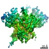
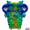
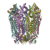
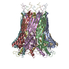
| Title | cryo-EM structure of human Pannexin 1 channel | ||||||
 Components Components | Pannexin-1 | ||||||
 Keywords Keywords | MEMBRANE PROTEIN / ion channel / molecular penetrate | ||||||
| Function / homology |  Function and homology information Function and homology informationElectric Transmission Across Gap Junctions / leak channel activity / positive regulation of interleukin-1 alpha production / bleb / wide pore channel activity / gap junction channel activity / gap junction / positive regulation of macrophage cytokine production / oogenesis / response to ATP ...Electric Transmission Across Gap Junctions / leak channel activity / positive regulation of interleukin-1 alpha production / bleb / wide pore channel activity / gap junction channel activity / gap junction / positive regulation of macrophage cytokine production / oogenesis / response to ATP / monoatomic cation transport / The NLRP3 inflammasome / positive regulation of interleukin-1 beta production / response to ischemia / calcium channel activity / calcium ion transport / actin filament binding / cell-cell signaling / scaffold protein binding / protease binding / transmembrane transporter binding / signaling receptor binding / endoplasmic reticulum membrane / structural molecule activity / endoplasmic reticulum / protein-containing complex / membrane / identical protein binding / plasma membraneSimilarity search - Function | ||||||
| Biological species |  Homo sapiens (human) Homo sapiens (human) | ||||||
| Method | ELECTRON MICROSCOPY / single particle reconstruction / cryo EM / Resolution: 3.2 Å | ||||||
 Authors Authors | Ronggui, Q. / Lili, D. / Jilin, Z. / Xuekui, Y. / Lei, W. / Shujia, Z. | ||||||
| Funding support |  China, 1items China, 1items
| ||||||
 Citation Citation | Journal: Cell Res / Year: 2020 Title: Cryo-EM structure of human heptameric Pannexin 1 channel. Authors: Ronggui Qu / Lili Dong / Jilin Zhang / Xuekui Yu / Lei Wang / Shujia Zhu / | ||||||
| History |
|
- Structure visualization
| Movie |
Movie viewer |
|---|---|
| Structure viewer | Molecule: MolmilJmol/JSmol |
- Downloads & links
-Download
| PDBx/mmCIF format | 6m02.cif.gz | 404.4 KB | Display | PDBx/mmCIF format |
|---|---|---|---|---|
| PDB format | pdb6m02.ent.gz | 345 KB | Display | PDB format |
| PDBx/mmJSON format | 6m02.json.gz | Tree view | PDBx/mmJSON format | |
| Others |  Other downloads Other downloads |
-Validation report
| Arichive directory | https://data.pdbj.org/pub/pdb/validation_reports/m0/6m02ftp://data.pdbj.org/pub/pdb/validation_reports/m0/6m02 | HTTPS FTP |
|---|
-Related structure data
| Related structure data | 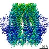 30028MC M: map data used to model this data C: citing same article ( |
|---|---|
| Similar structure data |


-Links
 PDBj
PDBj


- Assembly
| Deposited unit | 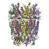
|
|---|---|
| 1 |
|
-Components
| #1: Protein | Mass: 48093.863 Da / Num. of mol.: 7 Source method: isolated from a genetically manipulated source Source: (gene. exp.) Homo sapiens (human) / Gene: PANX1, MRS1, UNQ2529/PRO6028 / Production host:  Eukaryota sp. (eukaryote) / References: UniProt: Q96RD7 Eukaryota sp. (eukaryote) / References: UniProt: Q96RD7 |
|---|
-Experimental details
-Experiment
| Experiment | Method: ELECTRON MICROSCOPY |
|---|---|
| EM experiment | Aggregation state: PARTICLE / 3D reconstruction method: single particle reconstruction |
- Sample preparation
| Component | Name: human pannexin protein 1 / Type: COMPLEX / Entity ID: all / Source: RECOMBINANT |
|---|---|
| Source (natural) | Organism: Homo sapiens (human) |
| Source (recombinant) | Organism: Eukaryota sp. (eukaryote) |
| Buffer solution | pH: 8 |
| Specimen | Embedding applied: NO / Shadowing applied: NO / Staining applied: NO / Vitrification applied: YES |
| Vitrification | Cryogen name: ETHANE |
- Electron microscopy imaging
| Experimental equipment |  Model: Titan Krios / Image courtesy: FEI Company |
|---|---|
| Microscopy | Model: FEI TITAN KRIOS |
| Electron gun | Electron source: FIELD EMISSION GUN / Accelerating voltage: 300 kV / Illumination mode: FLOOD BEAM |
| Electron lens | Mode: BRIGHT FIELDBright-field microscopy / Nominal magnification: 81000 X / Nominal defocus max: 3000 nm / Nominal defocus min: 1500 nm / Calibrated defocus min: 503 nm / Calibrated defocus max: 3574 nm |
| Image recording | Electron dose: 67 e/Å2 / Film or detector model: GATAN K3 (6k x 4k) |
- Processing
| CTF correction | Type: PHASE FLIPPING AND AMPLITUDE CORRECTION |
|---|---|
| Symmetry | Point symmetry: C7 (7 fold cyclic) |
| 3D reconstruction | Resolution: 3.2 Å / Resolution method: FSC 0.143 CUT-OFF / Num. of particles: 73421 / Symmetry type: POINT |
