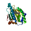
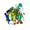
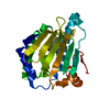
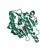
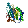
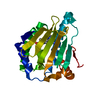
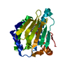
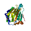
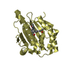
Heat shock protein HSP 90-alphaHeat shock response
Keywords
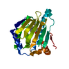
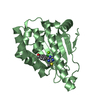
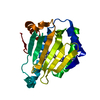
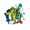
CHAPERONE / HSP90 / Ganetespib
Function / homology
Function and homology information
sperm mitochondrial sheath / dATP binding / Scavenging by Class F Receptors / sulfonylurea receptor binding / CTP binding / positive regulation of protein polymerization / vRNP Assembly / UTP binding / sperm plasma membrane / positive regulation of tau-protein kinase activity ...sperm mitochondrial sheath / dATP binding / Scavenging by Class F Receptors / sulfonylurea receptor binding / CTP binding / positive regulation of protein polymerization / vRNP Assembly / UTP binding / sperm plasma membrane / positive regulation of tau-protein kinase activity / protein insertion into mitochondrial outer membrane / chaperone-mediated autophagy / telomerase holoenzyme complex assembly / Rho GDP-dissociation inhibitor binding / Uptake and function of diphtheria toxin / mitochondrial transport / Drug-mediated inhibition of ERBB2 signaling / Resistance of ERBB2 KD mutants to trastuzumab / Resistance of ERBB2 KD mutants to sapitinib / Resistance of ERBB2 KD mutants to tesevatinib / Resistance of ERBB2 KD mutants to neratinib / Resistance of ERBB2 KD mutants to osimertinib / Resistance of ERBB2 KD mutants to afatinib / Resistance of ERBB2 KD mutants to AEE788 / Resistance of ERBB2 KD mutants to lapatinib / Drug resistance in ERBB2 TMD/JMD mutants / PIWI-interacting RNA (piRNA) biogenesis / TPR domain binding / non-chaperonin molecular chaperone ATPase / regulation of postsynaptic membrane neurotransmitter receptor levels / dendritic growth cone / Sema3A PAK dependent Axon repulsion / skeletal muscle contraction / regulation of protein ubiquitination / positive regulation of cell size / protein unfolding / HSF1-dependent transactivation / telomere maintenance via telomerase / response to unfolded protein / HSF1 activation / chaperone-mediated protein complex assembly / regulation of protein-containing complex assembly / Attenuation phase / RHOBTB2 GTPase cycle / positive regulation of lamellipodium assembly / eNOS activation / DNA polymerase binding / axonal growth cone / Tetrahydrobiopterin (BH4) synthesis, recycling, salvage and regulation / Loss of Nlp from mitotic centrosomes / Loss of proteins required for interphase microtubule organization from the centrosome / positive regulation of cardiac muscle contraction / Signaling by ERBB2 / cardiac muscle cell apoptotic process / Recruitment of mitotic centrosome proteins and complexes / positive regulation of telomerase activity / response to salt stress / positive regulation of defense response to virus by host / endocytic vesicle lumen / Recruitment of NuMA to mitotic centrosomes / HSP90 chaperone cycle for steroid hormone receptors (SHR) in the presence of ligand / Anchoring of the basal body to the plasma membrane / protein tyrosine kinase binding / response to cold / activation of innate immune response / positive regulation of interferon-beta production / nitric-oxide synthase regulator activity / lysosomal lumen / Constitutive Signaling by Overexpressed ERBB2 / ESR-mediated signaling / AURKA Activation by TPX2 / VEGFR2 mediated vascular permeability / response to cocaine / brush border membrane / ATP-dependent protein folding chaperone / Signaling by ERBB2 TMD/JMD mutants / neuron migration / Constitutive Signaling by EGFRvIII / DDX58/IFIH1-mediated induction of interferon-alpha/beta / Signaling by ERBB2 ECD mutants / tau protein binding / Signaling by ERBB2 KD Mutants / Regulation of necroptotic cell death / Regulation of actin dynamics for phagocytic cup formation / Downregulation of ERBB2 signaling / cellular response to virus / VEGFA-VEGFR2 Pathway / Aggrephagy / Chaperone Mediated Autophagy / positive regulation of protein import into nucleus / response to estrogen / histone deacetylase binding / positive regulation of protein catabolic process / The role of GTSE1 in G2/M progression after G2 checkpoint / regulation of protein localization / positive regulation of nitric oxide biosynthetic process / disordered domain specific binding / Regulation of PLK1 Activity at G2/M Transition / unfolded protein binding / melanosome Similarity search - Function
Heat shock protein Hsp90, conserved site / Heat shock hsp90 proteins family signature. / HSP90, C-terminal domain / Heat shock protein Hsp90, N-terminal / Heat shock protein Hsp90 family / Hsp90 protein / Histidine kinase-, DNA gyrase B-, and HSP90-like ATPase / Histidine kinase-, DNA gyrase B-, and HSP90-like ATPase / Histidine kinase-like ATPases / Histidine kinase/HSP90-like ATPase ...Heat shock protein Hsp90, conserved site / Heat shock hsp90 proteins family signature. / HSP90, C-terminal domain / Heat shock protein Hsp90, N-terminal / Heat shock protein Hsp90 family / Hsp90 protein / Histidine kinase-, DNA gyrase B-, and HSP90-like ATPase / Histidine kinase-, DNA gyrase B-, and HSP90-like ATPase / Histidine kinase-like ATPases / Histidine kinase/HSP90-like ATPase / Histidine kinase/HSP90-like ATPase superfamily / Ribosomal protein S5 domain 2-type fold Similarity search - Domain/homology
In the structure databanks used in Yorodumi, some data are registered as the other names, "COVID-19 virus" and "2019-nCoV". Here are the details of the virus and the list of structure data.
Jan 31, 2019. EMDB accession codes are about to change! (news from PDBe EMDB page)
EMDB accession codes are about to change! (news from PDBe EMDB page)
The allocation of 4 digits for EMDB accession codes will soon come to an end. Whilst these codes will remain in use, new EMDB accession codes will include an additional digit and will expand incrementally as the available range of codes is exhausted. The current 4-digit format prefixed with “EMD-” (i.e. EMD-XXXX) will advance to a 5-digit format (i.e. EMD-XXXXX), and so on. It is currently estimated that the 4-digit codes will be depleted around Spring 2019, at which point the 5-digit format will come into force.
The EM Navigator/Yorodumi systems omit the EMD- prefix.
Related info.:Q: What is EMD? / ID/Accession-code notation in Yorodumi/EM Navigator
Yorodumi is a browser for structure data from EMDB, PDB, SASBDB, etc.
This page is also the successor to EM Navigator detail page, and also detail information page/front-end page for Omokage search.
The word "yorodu" (or yorozu) is an old Japanese word meaning "ten thousand". "mi" (miru) is to see.
Related info.:EMDB / PDB / SASBDB / Comparison of 3 databanks / Yorodumi Search / Aug 31, 2016. New EM Navigator & Yorodumi / Yorodumi Papers / Jmol/JSmol / Function and homology information / Changes in new EM Navigator and Yorodumi
 Movie
Movie Controller


 Open data
 Basic information
 Components
 Heat shock response
  Keywords
 Function and homology information
 Authors
 Citation
 Structure visualization
 Downloads & links
 Other downloads
 PDBj
PDBj


 Assembly
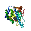

 Mass: 364.398 Da / Num. of mol.: 1 / Source method: isolated from a natural source / Formula: C20H20N4O3 / Feature type: SUBJECT OF INVESTIGATION
 Mass: 18.015 Da / Num. of mol.: 98 / Source method: isolated from a natural source / Formula: H2O
 Sample preparation
 / Beamline: BL17U1 / Wavelength: 0.979 Å
 Processing