 Movie
Movie Controller


+ Open data

- Basic information
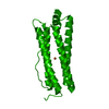
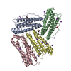
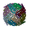
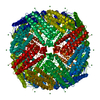
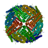
| Entry | Database: PDB / ID: 6lpd | ||||||
|---|---|---|---|---|---|---|---|
| Title | Phascolosoma esculenta | ||||||
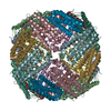
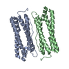
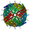
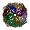
 Components Components | Ferritin | ||||||
 Keywords Keywords | OXIDOREDUCTASE / Phascolosoma esculenta (Fer147) / ferritin / STRUCTURAL PROTEIN | ||||||
| Function / homology | : / :  Function and homology information Function and homology information | ||||||
| Biological species |  Phascolosoma esculenta (invertebrata) Phascolosoma esculenta (invertebrata) | ||||||
| Method | X-RAY DIFFRACTION / SYNCHROTRON / MOLECULAR REPLACEMENT / Resolution: 1.65 Å | ||||||
 Authors Authors | Su, X.R. / Ming, T.H. | ||||||
| Funding support |  China, 1items China, 1items
| ||||||
 Citation Citation | Journal: Febs Open Bio / Year: 2021 Title: Structural comparison of two ferritins from the marine invertebrate Phascolosoma esculenta. Authors: Ming, T. / Huan, H. / Su, C. / Huo, C. / Wu, Y. / Jiang, Q. / Qiu, X. / Lu, C. / Zhou, J. / Li, Y. / Su, X. | ||||||
| History |
|
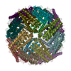
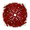
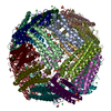
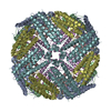
- Structure visualization
| Structure viewer | Molecule: MolmilJmol/JSmol |
|---|
- Downloads & links
-Download
| PDBx/mmCIF format | 6lpd.cif.gz | 518.1 KB | Display | PDBx/mmCIF format |
|---|---|---|---|---|
| PDB format | pdb6lpd.ent.gz | 429.7 KB | Display | PDB format |
| PDBx/mmJSON format | 6lpd.json.gz | Tree view | PDBx/mmJSON format | |
| Others |  Other downloads Other downloads |
-Validation report
| Arichive directory | https://data.pdbj.org/pub/pdb/validation_reports/lp/6lpdftp://data.pdbj.org/pub/pdb/validation_reports/lp/6lpd | HTTPS FTP |
|---|
-Related structure data
| Related structure data | 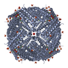 6lpeC  3ajoS S: Starting model for refinement C: citing same article ( |
|---|---|
| Similar structure data |


-Links
 PDBj
PDBj


- Assembly
| Deposited unit | 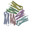
| ||||||||
|---|---|---|---|---|---|---|---|---|---|
| 1 | 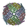
| ||||||||
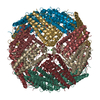
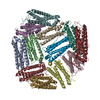
| Unit cell |
|
-Components
| #1: Protein | Mass: 20426.783 Da / Num. of mol.: 6 Source method: isolated from a genetically manipulated source Source: (gene. exp.) Phascolosoma esculenta (invertebrata) / Production host:  Escherichia coli BL21(DE3) (bacteria) / Strain (production host): BL21(DE3) Escherichia coli BL21(DE3) (bacteria) / Strain (production host): BL21(DE3)#2: Chemical | ChemComp-FE2 /   Mass: 55.845 Da / Num. of mol.: 13 / Source method: obtained synthetically / Formula: Fe / Feature type: SUBJECT OF INVESTIGATION Mass: 55.845 Da / Num. of mol.: 13 / Source method: obtained synthetically / Formula: Fe / Feature type: SUBJECT OF INVESTIGATION#3: Chemical | ChemComp-FE / | Iron  Mass: 55.845 Da / Num. of mol.: 1 / Source method: obtained synthetically / Formula: Fe / Feature type: SUBJECT OF INVESTIGATION Mass: 55.845 Da / Num. of mol.: 1 / Source method: obtained synthetically / Formula: Fe / Feature type: SUBJECT OF INVESTIGATION#4: Water | ChemComp-HOH / | Water Mass: 18.015 Da / Num. of mol.: 1459 / Source method: isolated from a natural source / Formula: H2O Mass: 18.015 Da / Num. of mol.: 1459 / Source method: isolated from a natural source / Formula: H2OHas ligand of interest | Y | |
|---|
-Experimental details
-Experiment
| Experiment | Method: X-RAY DIFFRACTION / Number of used crystals: 1 |
|---|
- Sample preparation
| Crystal grow | Temperature: 291.15 K / Method: vapor diffusion, sitting drop / pH: 7.5 / Details: 0.1 M HEPES pH 7.5, 20% v/v Jeffamine M-600 |
|---|
-Data collection
| Diffraction |
| |||||||||||||||
|---|---|---|---|---|---|---|---|---|---|---|---|---|---|---|---|---|
| Diffraction source |
| |||||||||||||||
| Detector |
| |||||||||||||||
| Radiation |
| |||||||||||||||
| Radiation wavelength |
| |||||||||||||||
| Reflection | Resolution: 1.65→108.65 Å / Num. obs: 215690 / % possible obs: 96.383 % / Redundancy: 5.3 % / CC1/2: 0.88 / Net I/σ(I): 38.3 | |||||||||||||||
| Reflection shell | Resolution: 1.65→1.66 Å / Num. unique obs: 10411 / CC1/2: 0.88 | |||||||||||||||
| Serial crystallography sample delivery |
|
- Processing
| Software |
| |||||||||||||||||||||||||||||||||||||||||||||||||||||||||||||||||||||||||||
|---|---|---|---|---|---|---|---|---|---|---|---|---|---|---|---|---|---|---|---|---|---|---|---|---|---|---|---|---|---|---|---|---|---|---|---|---|---|---|---|---|---|---|---|---|---|---|---|---|---|---|---|---|---|---|---|---|---|---|---|---|---|---|---|---|---|---|---|---|---|---|---|---|---|---|---|---|
| Refinement | Method to determine structure: MOLECULAR REPLACEMENT Starting model: 3AJO Resolution: 1.65→108.65 Å / Cor.coef. Fo:Fc: 0.974 / Cor.coef. Fo:Fc free: 0.954 / SU B: 1.913 / SU ML: 0.03 / Cross valid method: THROUGHOUT / σ(F): 0 / ESU R: 0.061 / ESU R Free: 0.061 / Stereochemistry target values: MAXIMUM LIKELIHOOD Details: HYDROGENS HAVE BEEN ADDED IN THE RIDING POSITIONS U VALUES : REFINED INDIVIDUALLY
| |||||||||||||||||||||||||||||||||||||||||||||||||||||||||||||||||||||||||||
| Solvent computation | Ion probe radii: 0.8 Å / Shrinkage radii: 0.8 Å / VDW probe radii: 1.2 Å / Solvent model: MASK | |||||||||||||||||||||||||||||||||||||||||||||||||||||||||||||||||||||||||||
| Displacement parameters | Biso max: 150.46 Å2 / Biso mean: 17.279 Å2 / Biso min: 5.64 Å2
| |||||||||||||||||||||||||||||||||||||||||||||||||||||||||||||||||||||||||||
| Refinement step | Cycle: final / Resolution: 1.65→108.65 Å
| |||||||||||||||||||||||||||||||||||||||||||||||||||||||||||||||||||||||||||
| Refine LS restraints |
| |||||||||||||||||||||||||||||||||||||||||||||||||||||||||||||||||||||||||||
| LS refinement shell | Resolution: 1.65→1.693 Å / Rfactor Rfree error: 0 / Total num. of bins used: 20
|
