 Movie
Movie Controller

[English] 日本語
 Yorodumi
- PDB-6lp3: Structural basis and functional analysis epo1-bem3p complex for b... -
+ Open data

- Basic information
| Entry | Database: PDB / ID: 6lp3 | ||||||
|---|---|---|---|---|---|---|---|
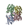
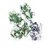
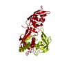
| Title | Structural basis and functional analysis epo1-bem3p complex for bud growth | ||||||
 Components Components |
| ||||||
 Keywords Keywords |  CELL ADHESION / Budding yeast / Organelle inheritance / Endoplasmic reticulum inheritance / Polarisome / Epo1p / Bem3p. CELL ADHESION / Budding yeast / Organelle inheritance / Endoplasmic reticulum inheritance / Polarisome / Epo1p / Bem3p. | ||||||
| Function / homology |  Function and homology information Function and homology informationendoplasmic reticulum polarization / : / : / RHOF GTPase cycle / CDC42 GTPase cycle / RHOD GTPase cycle / RHOV GTPase cycle / septin ring organization / RHOA GTPase cycle / incipient cellular bud site ...endoplasmic reticulum polarization / : / : / RHOF GTPase cycle / CDC42 GTPase cycle / RHOD GTPase cycle / RHOV GTPase cycle / septin ring organization / RHOA GTPase cycle / incipient cellular bud site / cellular bud tip / cellular bud neck / mating projection tip / phosphatidylinositol-3-phosphate binding / negative regulation of Rho protein signal transduction / establishment of cell polarity / regulation of GTPase activity / Neutrophil degranulation / GTPase activator activity / positive regulation of GTPase activity / cell cortex / signal transduction / cytoplasmSimilarity search - Function | ||||||
| Biological species |  Saccharomyces cerevisiae (brewer's yeast) Saccharomyces cerevisiae (brewer's yeast) | ||||||
| Method | X-RAY DIFFRACTION / SYNCHROTRON / MAD / Resolution: 3.547 Å | ||||||
 Authors Authors | Wang, J. / Li, L. / Ming, Z.H. / Wu, L.J. / Yan, L.M. | ||||||
 Citation Citation | Journal: To Be Published Title: Structural basis and functional analysis epo1-bem3p complex for bud growth Authors: Wang, J. | ||||||
| History |
|
- Structure visualization
| Structure viewer | Molecule: MolmilJmol/JSmol |
|---|
- Downloads & links
-Download
| PDBx/mmCIF format | 6lp3.cif.gz | 130.7 KB | Display | PDBx/mmCIF format |
|---|---|---|---|---|
| PDB format | pdb6lp3.ent.gz | 101.2 KB | Display | PDB format |
| PDBx/mmJSON format | 6lp3.json.gz | Tree view | PDBx/mmJSON format | |
| Others |  Other downloads Other downloads |
-Validation report
| Arichive directory | https://data.pdbj.org/pub/pdb/validation_reports/lp/6lp3ftp://data.pdbj.org/pub/pdb/validation_reports/lp/6lp3 | HTTPS FTP |
|---|
-Related structure data


-Links
 PDBj
PDBj


- Assembly
| Deposited unit | 
| ||||||||
|---|---|---|---|---|---|---|---|---|---|
| 1 |
| ||||||||
| Unit cell |
|
-Components
| #1: Protein | Mass: 22375.828 Da / Num. of mol.: 4 Source method: isolated from a genetically manipulated source Source: (gene. exp.) Saccharomyces cerevisiae (strain ATCC 204508 / S288c) (yeast)Strain: ATCC 204508 / S288c / Gene: YMR124W, YM8564.06 / Production host:  Escherichia coli (E. coli) / References: UniProt: P39523 Escherichia coli (E. coli) / References: UniProt: P39523#2: Protein | Mass: 11400.604 Da / Num. of mol.: 2 Source method: isolated from a genetically manipulated source Source: (gene. exp.) Saccharomyces cerevisiae (strain ATCC 204508 / S288c) (yeast)Strain: ATCC 204508 / S288c / Gene: BEM3, YPL115C, LPH12C / Production host: Escherichia coli (E. coli) / References: UniProt: P32873 |
|---|
-Experimental details
-Experiment
| Experiment | Method: X-RAY DIFFRACTION / Number of used crystals: 1 |
|---|
- Sample preparation
| Crystal | Density Matthews: 2.29 Å3/Da / Density % sol: 46.39 % |
|---|---|
| Crystal grow | Temperature: 289 K / Method: vapor diffusion, hanging drop / pH: 6.5 Details: 1.6 M Ammonium sulfate, 0.1 M MES, pH 6.5, 10% 1,4-Dioxane |
-Data collection
| Diffraction | Mean temperature: 100 K / Serial crystal experiment: N |
|---|---|
| Diffraction source | Source: SYNCHROTRON / Site: SSRF  / Beamline: BL17U / Wavelength: 0.9789 Å / Beamline: BL17U / Wavelength: 0.9789 Å |
| Detector | Type: ADSC QUANTUM 315 / Detector: CCD / Date: Jun 12, 2018 |
| Radiation | Protocol: SINGLE WAVELENGTH / Monochromatic (M) / Laue (L): M / Scattering type: x-ray |
| Radiation wavelength | Wavelength: 0.9789 Å / Relative weight: 1 |
| Reflection | Resolution: 3.547→46.032 Å / Num. obs: 13161 / % possible obs: 99.9 % / Redundancy: 18 % / Rmerge(I) obs: 0.431 / Net I/σ(I): 10.31 |
| Reflection shell | Resolution: 3.95→4.23 Å / Rmerge(I) obs: 0.961 / Num. unique obs: 1249 |
- Processing
| Software |
| ||||||||||||||||||||||||||||||||||||
|---|---|---|---|---|---|---|---|---|---|---|---|---|---|---|---|---|---|---|---|---|---|---|---|---|---|---|---|---|---|---|---|---|---|---|---|---|---|
| Refinement | Method to determine structure: MAD / Resolution: 3.547→46.032 Å / SU ML: 0.68 / Cross valid method: THROUGHOUT / σ(F): 1.34 / Phase error: 46.48 / Stereochemistry target values: ML
| ||||||||||||||||||||||||||||||||||||
| Solvent computation | Shrinkage radii: 0.9 Å / VDW probe radii: 1.11 Å / Solvent model: FLAT BULK SOLVENT MODEL | ||||||||||||||||||||||||||||||||||||
| Displacement parameters | Biso max: 383.6 Å2 / Biso mean: 122.3199 Å2 / Biso min: 30 Å2 | ||||||||||||||||||||||||||||||||||||
| Refinement step | Cycle: final / Resolution: 3.547→46.032 Å
| ||||||||||||||||||||||||||||||||||||
| LS refinement shell | Refine-ID: X-RAY DIFFRACTION / Rfactor Rfree error: 0
|
