 Movie
Movie Controller

[English] 日本語
 Yorodumi
- PDB-6lm3: Neutralization mechanism of a monoclonal antibody targeting a por... -
+ Open data

- Basic information
| Entry | Database: PDB / ID: 6lm3 | ||||||
|---|---|---|---|---|---|---|---|
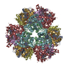
| Title | Neutralization mechanism of a monoclonal antibody targeting a porcine circovirus type 2 Cap protein conformational epitope | ||||||
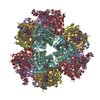
 Components Components | Capsid protein Capsid Capsid | ||||||
 Keywords Keywords | VIRUS / Porcine circovirus type 2 / Cap protein | ||||||
| Function / homology | Circovirus capsid protein / Circovirus capsid superfamily / Circovirus capsid protein / viral capsid assembly / T=1 icosahedral viral capsid / symbiont entry into host cell / virion attachment to host cell / Capsid protein Function and homology information Function and homology information | ||||||
| Biological species |  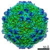 Porcine circovirus 2 Porcine circovirus 2 | ||||||
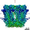
| Method | ELECTRON MICROSCOPY / single particle reconstruction / cryo EM / Resolution: 6.7 Å | ||||||
 Authors Authors | Sun, Z. / Huang, L. / Xia, D. / Wei, Y. / Sun, E. / Zhu, H. / Bian, H. / Wu, H. / Feng, L. / Wang, J. / Liu, C. | ||||||
| Funding support |  China, 1items China, 1items
| ||||||
 Citation Citation | Journal: J Virol / Year: 2020 Title: Neutralization Mechanism of a Monoclonal Antibody Targeting a Porcine Circovirus Type 2 Cap Protein Conformational Epitope. Authors: Liping Huang / Zhenzhao Sun / Deli Xia / Yanwu Wei / Encheng Sun / Chunguo Liu / Hongzhen Zhu / Haiqiao Bian / Hongli Wu / Li Feng / Jingfei Wang / Changming Liu / Abstract: Porcine circovirus type 2 (PCV2) is an important pathogen in swine herds, and its infection of pigs has caused severe economic losses to the pig industry worldwide. The capsid protein of PCV2 is the ...Porcine circovirus type 2 (PCV2) is an important pathogen in swine herds, and its infection of pigs has caused severe economic losses to the pig industry worldwide. The capsid protein of PCV2 is the only structural protein that is associated with PCV2 infection and immunity. Here, we report a neutralizing monoclonal antibody (MAb), MAb 3A5, that binds to intact PCV2 virions of the PCV2a, PCV2b, and PCV2d genotypes. MAb 3A5 neutralized PCV2 by blocking viral attachment to PK15 cells. To further explore the neutralization mechanism, we resolved the structure of the PCV2 virion in complex with MAb 3A5 Fab fragments by using cryo-electron microscopy single-particle analysis. The binding sites were located at the topmost edges around 5-fold icosahedral symmetry axes, with each footprint covering amino acids from two adjacent capsid proteins. Most of the epitope residues (15/18 residues) were conserved among 2,273 PCV2 strains. Mutations of some amino acids within the epitope had significant effects on the neutralizing activity of MAb 3A5. This study reveals the molecular and structural bases of this PCV2-neutralizing antibody and provides new and important information for vaccine design and therapeutic antibody development against PCV2 infections. PCV2 is associated with several clinical manifestations collectively known as PCV2-associated diseases (PCVADs). Neutralizing antibodies play a crucial role in the prevention of PCVADs. We demonstrated previously that a MAb, MAb 3A5, neutralizes the PCV2a, PCV2b, and PCV2d genotypes with different degrees of efficiency, but the underlying mechanism remains elusive. Here, we report the neutralization mechanism of this MAb and the structure of the PCV2 virion in complex with MAb 3A5 Fabs, showing a binding mode in which one Fab interacted with more than two loops from two adjacent capsid proteins. This binding mode has not been observed previously for PCV2-neutralizing antibodies. Our work provides new and important information for vaccine design and therapeutic antibody development against PCV2 infections. | ||||||
| History |
|
- Structure visualization
| Movie |
Movie viewer |
|---|---|
| Structure viewer | Molecule: MolmilJmol/JSmol |
- Downloads & links
-Download
| PDBx/mmCIF format | 6lm3.cif.gz | 48.7 KB | Display | PDBx/mmCIF format |
|---|---|---|---|---|
| PDB format | pdb6lm3.ent.gz | 32.3 KB | Display | PDB format |
| PDBx/mmJSON format | 6lm3.json.gz | Tree view | PDBx/mmJSON format | |
| Others |  Other downloads Other downloads |
-Validation report
| Arichive directory | https://data.pdbj.org/pub/pdb/validation_reports/lm/6lm3ftp://data.pdbj.org/pub/pdb/validation_reports/lm/6lm3 | HTTPS FTP |
|---|
-Related structure data
| Related structure data | 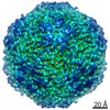 0916MC  0838C 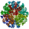 6l62C M: map data used to model this data C: citing same article ( |
|---|---|
| Similar structure data |


-Links
 PDBj
PDBj
- Assembly
| Deposited unit | 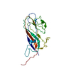
|
|---|---|
| 1 | x 60
|
| 2 |
|
| 3 | x 5
|
| 4 | x 6
|
| 5 | 
|
| Symmetry | Point symmetry: (Schoenflies symbol: I (icosahedral)) |
-Components
| #1: Protein | Capsid Mass: 27559.520 Da / Num. of mol.: 1 Source method: isolated from a genetically manipulated source Source: (gene. exp.) Porcine circovirus 2 / Production host: Porcine circovirus 2 / References: UniProt: F5A4Z4 |
|---|
-Experimental details
-Experiment
| Experiment | Method: ELECTRON MICROSCOPY |
|---|---|
| EM experiment | Aggregation state: PARTICLE / 3D reconstruction method: single particle reconstruction |
- Sample preparation
| Component | Name: PCV2 virion in complex with Fab fragments of the mAb 3A5 Type: COMPLEX / Entity ID: all / Source: NATURAL |
|---|---|
| Source (natural) | Organism: Porcine circovirus 2 |
| Buffer solution | pH: 7.4 |
| Specimen | Embedding applied: NO / Shadowing applied: NO / Staining applied: NO / Vitrification applied: YES |
| Vitrification | Cryogen name: ETHANE |
- Electron microscopy imaging
| Experimental equipment |  Model: Talos Arctica / Image courtesy: FEI Company |
|---|---|
| Microscopy | Model: FEI TALOS ARCTICA |
| Electron gun | Electron source: FIELD EMISSION GUN / Accelerating voltage: 200 kV / Illumination mode: FLOOD BEAM |
| Electron lens | Mode: BRIGHT FIELDBright-field microscopy |
| Image recording | Electron dose: 35 e/Å2 / Film or detector model: FEI CETA (4k x 4k) |
- Processing
| Software |
| ||||||||||||||||||||||||
|---|---|---|---|---|---|---|---|---|---|---|---|---|---|---|---|---|---|---|---|---|---|---|---|---|---|
| CTF correction | Type: NONE | ||||||||||||||||||||||||
| 3D reconstruction | Resolution: 6.7 Å / Resolution method: FSC 0.143 CUT-OFF / Num. of particles: 2011 / Symmetry type: POINT | ||||||||||||||||||||||||
| Refinement | Stereochemistry target values: GeoStd + Monomer Library | ||||||||||||||||||||||||
| Refine LS restraints |
|
