 Movie
Movie Controller


+ Open data

- Basic information
| Entry | Database: PDB / ID: 6lko | |||||||||
|---|---|---|---|---|---|---|---|---|---|---|
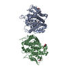
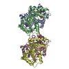
| Title | Turning an asparaginyl endopeptidase into a peptide ligase | |||||||||
 Components Components | Asparaginyl endopeptidase Asparagine endopeptidase Asparagine endopeptidase | |||||||||
 Keywords Keywords | HYDROLASE / AEP / Asparaginyl Endopeptidase / Peptide asparaginyl Ligase / PAL | |||||||||
| Function / homology |  Function and homology informationlegumain / proteolysis involved in protein catabolic process / cysteine-type endopeptidase activity Function and homology informationlegumain / proteolysis involved in protein catabolic process / cysteine-type endopeptidase activitySimilarity search - Function | |||||||||
| Biological species |  Clitoria ternatea (plant) Clitoria ternatea (plant) | |||||||||
| Method | X-RAY DIFFRACTION / SYNCHROTRON / MOLECULAR REPLACEMENT / Resolution: 2 Å | |||||||||
 Authors Authors | El Sahili, A. / Lescar, J. | |||||||||
| Funding support |  Singapore, 1items Singapore, 1items
| |||||||||
 Citation Citation | Journal: Acs Catalysis / Year: 2020 Title: Turning an Asparaginyl Endopeptidase into a Peptide Ligase Authors: Hemu, X. / El Sahili, A. / Hu, S. / Zhang, X. / Serra, A. / Goh, B.C. / Darwis, D.A. / Chen, M.W. / Sze, S.K. / Liu, C.F. / Lescar, J. / Tam, J.P. | |||||||||
| History |
|
- Structure visualization
| Structure viewer | Molecule: MolmilJmol/JSmol |
|---|
- Downloads & links
-Download
| PDBx/mmCIF format | 6lko.cif.gz | 115.3 KB | Display | PDBx/mmCIF format |
|---|---|---|---|---|
| PDB format | pdb6lko.ent.gz | 77.2 KB | Display | PDB format |
| PDBx/mmJSON format | 6lko.json.gz | Tree view | PDBx/mmJSON format | |
| Others |  Other downloads Other downloads |
-Validation report
| Arichive directory | https://data.pdbj.org/pub/pdb/validation_reports/lk/6lkoftp://data.pdbj.org/pub/pdb/validation_reports/lk/6lko | HTTPS FTP |
|---|
-Related structure data
| Related structure data |  6l4vSC  6l4wC  6l4xC  6l4yC S: Starting model for refinement C: citing same article ( |
|---|---|
| Similar structure data |


-Links
 PDBj
PDBj
- Assembly
| Deposited unit | 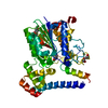
| ||||||||||||
|---|---|---|---|---|---|---|---|---|---|---|---|---|---|
| 1 | 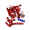
| ||||||||||||
| Unit cell |
|
-Components
| #1: Protein | Asparagine endopeptidase Mass: 55789.918 Da / Num. of mol.: 1 / Mutation: P183A, G252V Source method: isolated from a genetically manipulated source Source: (gene. exp.) Clitoria ternatea (plant) / Production host:   Spodoptera frugiperda (fall armyworm) / References: UniProt: A0A0P0QM28, legumain Spodoptera frugiperda (fall armyworm) / References: UniProt: A0A0P0QM28, legumain |
|---|---|
| #2: Sugar | ChemComp-NAG / N-Acetylglucosamine  Type: D-saccharide, beta linking / Mass: 221.208 Da / Num. of mol.: 1 Type: D-saccharide, beta linking / Mass: 221.208 Da / Num. of mol.: 1Source method: isolated from a genetically manipulated source Formula: C8H15NO6 |
| #3: Chemical | ChemComp-PEG / Diethylene glycol  Mass: 106.120 Da / Num. of mol.: 1 / Source method: obtained synthetically / Formula: C4H10O3 Mass: 106.120 Da / Num. of mol.: 1 / Source method: obtained synthetically / Formula: C4H10O3 |
| #4: Water | ChemComp-HOH / Water Mass: 18.015 Da / Num. of mol.: 288 / Source method: isolated from a natural source / Formula: H2O Mass: 18.015 Da / Num. of mol.: 288 / Source method: isolated from a natural source / Formula: H2O |
| Has ligand of interest | N |
-Experimental details
-Experiment
| Experiment | Method: X-RAY DIFFRACTION / Number of used crystals: 1 |
|---|
- Sample preparation
| Crystal | Density Matthews: 2.15 Å3/Da / Density % sol: 42.75 % |
|---|---|
| Crystal grow | Temperature: 293 K / Method: vapor diffusion, sitting drop / pH: 7.5 Details: 20% v/v PEG 500* MME, 10 % w/v PEG 20000, 0.1M Sodium formate, 0.1M Ammonium acetate, 0.1M Sodium citrate tribasic dihydrate, 0.1M Potassium sodium tartrate tetrahydrate, 0.1M Sodium ...Details: 20% v/v PEG 500* MME, 10 % w/v PEG 20000, 0.1M Sodium formate, 0.1M Ammonium acetate, 0.1M Sodium citrate tribasic dihydrate, 0.1M Potassium sodium tartrate tetrahydrate, 0.1M Sodium oxamate, 0.1M Sodium HEPES, MOPS (acid) |
-Data collection
| Diffraction | Mean temperature: 100 K / Serial crystal experiment: N |
|---|---|
| Diffraction source | Source: SYNCHROTRON / Site: SLS  / Beamline: X06DA / Wavelength: 1.00003 Å / Beamline: X06DA / Wavelength: 1.00003 Å |
| Detector | Type: DECTRIS PILATUS 2M-F / Detector: PIXEL / Date: Nov 10, 2019 |
| Radiation | Protocol: SINGLE WAVELENGTH / Monochromatic (M) / Laue (L): M / Scattering type: x-ray |
| Radiation wavelength | Wavelength: 1.00003 Å / Relative weight: 1 |
| Reflection | Resolution: 2→42.31 Å / Num. obs: 60392 / % possible obs: 99.96 % / Redundancy: 13 % / Biso Wilson estimate: 32.12 Å2 / CC1/2: 1 / Rmerge(I) obs: 0.093 / Net I/σ(I): 22.66 |
| Reflection shell | Resolution: 2→2.07 Å / Rmerge(I) obs: 1.076 / Num. unique obs: 3230 / CC1/2: 0.784 |
- Processing
| Software |
| |||||||||||||||||||||||||||||||||||||||||||||||||||||||||||||||||||||||||||||||||||||||||||||||||||||||||||||||||||||||||||||||||||||||||||||||||||||||||||||||||
|---|---|---|---|---|---|---|---|---|---|---|---|---|---|---|---|---|---|---|---|---|---|---|---|---|---|---|---|---|---|---|---|---|---|---|---|---|---|---|---|---|---|---|---|---|---|---|---|---|---|---|---|---|---|---|---|---|---|---|---|---|---|---|---|---|---|---|---|---|---|---|---|---|---|---|---|---|---|---|---|---|---|---|---|---|---|---|---|---|---|---|---|---|---|---|---|---|---|---|---|---|---|---|---|---|---|---|---|---|---|---|---|---|---|---|---|---|---|---|---|---|---|---|---|---|---|---|---|---|---|---|---|---|---|---|---|---|---|---|---|---|---|---|---|---|---|---|---|---|---|---|---|---|---|---|---|---|---|---|---|---|---|---|
| Refinement | Method to determine structure: MOLECULAR REPLACEMENT Starting model: 6L4V Resolution: 2→42.31 Å / SU ML: 0.2248 / Cross valid method: FREE R-VALUE / σ(F): 1.32 / Phase error: 21.3534 Stereochemistry target values: GeoStd + Monomer Library + CDL v1.2
| |||||||||||||||||||||||||||||||||||||||||||||||||||||||||||||||||||||||||||||||||||||||||||||||||||||||||||||||||||||||||||||||||||||||||||||||||||||||||||||||||
| Solvent computation | Shrinkage radii: 0.9 Å / VDW probe radii: 1.11 Å / Solvent model: FLAT BULK SOLVENT MODEL | |||||||||||||||||||||||||||||||||||||||||||||||||||||||||||||||||||||||||||||||||||||||||||||||||||||||||||||||||||||||||||||||||||||||||||||||||||||||||||||||||
| Displacement parameters | Biso mean: 37.4 Å2 | |||||||||||||||||||||||||||||||||||||||||||||||||||||||||||||||||||||||||||||||||||||||||||||||||||||||||||||||||||||||||||||||||||||||||||||||||||||||||||||||||
| Refinement step | Cycle: LAST / Resolution: 2→42.31 Å
| |||||||||||||||||||||||||||||||||||||||||||||||||||||||||||||||||||||||||||||||||||||||||||||||||||||||||||||||||||||||||||||||||||||||||||||||||||||||||||||||||
| Refine LS restraints |
| |||||||||||||||||||||||||||||||||||||||||||||||||||||||||||||||||||||||||||||||||||||||||||||||||||||||||||||||||||||||||||||||||||||||||||||||||||||||||||||||||
| LS refinement shell |
|
