 Movie
Movie Controller


+ Open data

- Basic information
| Entry | Database: PDB / ID: 6lj9 | ||||||||||||
|---|---|---|---|---|---|---|---|---|---|---|---|---|---|
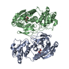
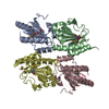
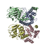
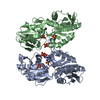
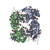
| Title | Crystal Structure of Se-Met ASFV pS273R protease | ||||||||||||
 Components Components | Cysteine protease S273R | ||||||||||||
 Keywords Keywords |  HYDROLASE HYDROLASE | ||||||||||||
| Function / homology |  Function and homology informationvirion component / host cell cytoplasm / Hydrolases; Acting on peptide bonds (peptidases); Cysteine endopeptidases / viral protein processing / cysteine-type endopeptidase activity / proteolysis Function and homology informationvirion component / host cell cytoplasm / Hydrolases; Acting on peptide bonds (peptidases); Cysteine endopeptidases / viral protein processing / cysteine-type endopeptidase activity / proteolysisSimilarity search - Function | ||||||||||||
| Biological species |  African swine fever virus pig/Kenya/KEN-50/1950 African swine fever virus pig/Kenya/KEN-50/1950 | ||||||||||||
| Method | X-RAY DIFFRACTION / SYNCHROTRON / SAD / Resolution: 2.307 Å | ||||||||||||
 Authors Authors | Li, G.B. / Liu, X.X. / Chen, C. / Guo, Y. | ||||||||||||
| Funding support |  China, 3items China, 3items
| ||||||||||||
 Citation Citation | Journal: J.Virol. / Year: 2020 Title: Crystal Structure of African Swine Fever Virus pS273R Protease and Implications for Inhibitor Design. Authors: Li, G. / Liu, X. / Yang, M. / Zhang, G. / Wang, Z. / Guo, K. / Gao, Y. / Jiao, P. / Sun, J. / Chen, C. / Wang, H. / Deng, W. / Xiao, H. / Li, S. / Wu, H. / Wang, Y. / Cao, L. / Jia, Z. / ...Authors: Li, G. / Liu, X. / Yang, M. / Zhang, G. / Wang, Z. / Guo, K. / Gao, Y. / Jiao, P. / Sun, J. / Chen, C. / Wang, H. / Deng, W. / Xiao, H. / Li, S. / Wu, H. / Wang, Y. / Cao, L. / Jia, Z. / Shang, L. / Yang, C. / Guo, Y. / Rao, Z. | ||||||||||||
| History |
|
- Structure visualization
| Structure viewer | Molecule: MolmilJmol/JSmol |
|---|
- Downloads & links
-Download
| PDBx/mmCIF format | 6lj9.cif.gz | 224.7 KB | Display | PDBx/mmCIF format |
|---|---|---|---|---|
| PDB format | pdb6lj9.ent.gz | 187.5 KB | Display | PDB format |
| PDBx/mmJSON format | 6lj9.json.gz | Tree view | PDBx/mmJSON format | |
| Others |  Other downloads Other downloads |
-Validation report
| Arichive directory | https://data.pdbj.org/pub/pdb/validation_reports/lj/6lj9ftp://data.pdbj.org/pub/pdb/validation_reports/lj/6lj9 | HTTPS FTP |
|---|
-Related structure data


-Links
 PDBj
PDBj- Assembly
| Deposited unit | 
| ||||||||
|---|---|---|---|---|---|---|---|---|---|
| 1 |
| ||||||||
| Unit cell |
|
-Components
| #1: Protein | Mass: 32915.051 Da / Num. of mol.: 2 Source method: isolated from a genetically manipulated source Source: (gene. exp.) African swine fever virus pig/Kenya/KEN-50/1950Strain: isolate Pig/Kenya/KEN-50/1950 / Gene: Ken-123 / Production host:  Escherichia coli (E. coli) Escherichia coli (E. coli)References: UniProt: P0C9B9, Hydrolases; Acting on peptide bonds (peptidases); Cysteine endopeptidases#2: Water | ChemComp-HOH / | Water Mass: 18.015 Da / Num. of mol.: 108 / Source method: isolated from a natural source / Formula: H2O Mass: 18.015 Da / Num. of mol.: 108 / Source method: isolated from a natural source / Formula: H2OHas ligand of interest | N | Sequence details | Authors states that they modeled the MSE residues (114A, 115A) in the place with a good quality ...Authors states that they modeled the MSE residues (114A, 115A) in the place with a good quality electron density. Thus, there have both the Met and Mse residues in the coordinates. | |
|---|
-Experimental details
-Experiment
| Experiment | Method: X-RAY DIFFRACTION / Number of used crystals: 1 |
|---|
- Sample preparation
| Crystal | Density Matthews: 2.09 Å3/Da / Density % sol: 41.14 % |
|---|---|
| Crystal grow | Temperature: 289 K / Method: vapor diffusion, sitting drop / pH: 8.2 Details: 0.2 M Lithium sulfate monohydrate, 0.1 M Tris, pH 8.2, 23% w/v Polyethylene glycol 1,500 PH range: 8.0-8.6 |
-Data collection
| Diffraction | Mean temperature: 100 K / Serial crystal experiment: N |
|---|---|
| Diffraction source | Source: SYNCHROTRON / Site: SPring-8  / Beamline: BL41XU / Wavelength: 0.97911 Å / Beamline: BL41XU / Wavelength: 0.97911 Å |
| Detector | Type: DECTRIS PILATUS3 6M / Detector: PIXEL / Date: Apr 17, 2019 |
| Radiation | Protocol: SINGLE WAVELENGTH / Monochromatic (M) / Laue (L): M / Scattering type: x-ray |
| Radiation wavelength | Wavelength: 0.97911 Å / Relative weight: 1 |
| Reflection | Resolution: 2.079→48.843 Å / Num. obs: 21872 / % possible obs: 100 % / Redundancy: 7.04 % / CC1/2: 0.999 / Net I/σ(I): 10.67 |
| Reflection shell | Resolution: 2.079→2.2 Å / Num. unique obs: 19824 / CC1/2: 0.715 |
- Processing
| Software |
| ||||||||||||||||||||||||||||||||||||||||||||||||||||||||||||||||||||||||||||||||||||||||||||||||||||||||||||||||||||||||||||||||||||||||||||||||||||||||||||||||||||||||||||||||||||||||||||||||||||||||
|---|---|---|---|---|---|---|---|---|---|---|---|---|---|---|---|---|---|---|---|---|---|---|---|---|---|---|---|---|---|---|---|---|---|---|---|---|---|---|---|---|---|---|---|---|---|---|---|---|---|---|---|---|---|---|---|---|---|---|---|---|---|---|---|---|---|---|---|---|---|---|---|---|---|---|---|---|---|---|---|---|---|---|---|---|---|---|---|---|---|---|---|---|---|---|---|---|---|---|---|---|---|---|---|---|---|---|---|---|---|---|---|---|---|---|---|---|---|---|---|---|---|---|---|---|---|---|---|---|---|---|---|---|---|---|---|---|---|---|---|---|---|---|---|---|---|---|---|---|---|---|---|---|---|---|---|---|---|---|---|---|---|---|---|---|---|---|---|---|---|---|---|---|---|---|---|---|---|---|---|---|---|---|---|---|---|---|---|---|---|---|---|---|---|---|---|---|---|---|---|---|---|
| Refinement | Method to determine structure: SAD / Resolution: 2.307→48.769 Å / SU ML: 0.24 / Cross valid method: THROUGHOUT / σ(F): 1.37 / Phase error: 22.97 / Stereochemistry target values: ML
| ||||||||||||||||||||||||||||||||||||||||||||||||||||||||||||||||||||||||||||||||||||||||||||||||||||||||||||||||||||||||||||||||||||||||||||||||||||||||||||||||||||||||||||||||||||||||||||||||||||||||
| Solvent computation | Shrinkage radii: 0.9 Å / VDW probe radii: 1.11 Å / Solvent model: FLAT BULK SOLVENT MODEL | ||||||||||||||||||||||||||||||||||||||||||||||||||||||||||||||||||||||||||||||||||||||||||||||||||||||||||||||||||||||||||||||||||||||||||||||||||||||||||||||||||||||||||||||||||||||||||||||||||||||||
| Displacement parameters | Biso max: 198.15 Å2 / Biso mean: 47.3544 Å2 / Biso min: 9.04 Å2 | ||||||||||||||||||||||||||||||||||||||||||||||||||||||||||||||||||||||||||||||||||||||||||||||||||||||||||||||||||||||||||||||||||||||||||||||||||||||||||||||||||||||||||||||||||||||||||||||||||||||||
| Refinement step | Cycle: final / Resolution: 2.307→48.769 Å
| ||||||||||||||||||||||||||||||||||||||||||||||||||||||||||||||||||||||||||||||||||||||||||||||||||||||||||||||||||||||||||||||||||||||||||||||||||||||||||||||||||||||||||||||||||||||||||||||||||||||||
| LS refinement shell | Refine-ID: X-RAY DIFFRACTION / Rfactor Rfree error: 0
| ||||||||||||||||||||||||||||||||||||||||||||||||||||||||||||||||||||||||||||||||||||||||||||||||||||||||||||||||||||||||||||||||||||||||||||||||||||||||||||||||||||||||||||||||||||||||||||||||||||||||
| Refinement TLS params. | Method: refined / Refine-ID: X-RAY DIFFRACTION
| ||||||||||||||||||||||||||||||||||||||||||||||||||||||||||||||||||||||||||||||||||||||||||||||||||||||||||||||||||||||||||||||||||||||||||||||||||||||||||||||||||||||||||||||||||||||||||||||||||||||||
| Refinement TLS group |
|
