 Movie
Movie Controller

[English] 日本語
 Yorodumi
- PDB-6lik: Crystal Structure of Lectin from Pleurotus ostreatus in complex w... -
+ Open data

- Basic information
| Entry | Database: PDB / ID: 6lik | ||||||
|---|---|---|---|---|---|---|---|
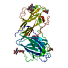
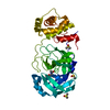
| Title | Crystal Structure of Lectin from Pleurotus ostreatus in complex with Galactose | ||||||
 Components Components | Lectin | ||||||
 Keywords Keywords | SUGAR BINDING PROTEIN / Pleurotus ostreatus Lectin / Mushroom Lectin / C-type lectin / Calcium mediated sugar binding | ||||||
| Function / homology | metal ion binding / alpha-D-galactopyranose / Lectin Function and homology information Function and homology information | ||||||
| Biological species |  Pleurotus ostreatus (oyster mushroom) Pleurotus ostreatus (oyster mushroom) | ||||||
| Method | X-RAY DIFFRACTION / SYNCHROTRON / MOLECULAR REPLACEMENT / molecular replacement / Resolution: 2.4 Å | ||||||
 Authors Authors | Vajravijayan, S. / Pletnev, S. / Luo, Z. / Iqbal, S. / Nandhagopal, N. / Gunasekaran, K. | ||||||
 Citation Citation | Journal: Int.J.Biol.Macromol. / Year: 2020 Title: Crystallographic and calorimetric analysis on Pleurotus ostreatus lectin and its sugar complexes - promiscuous binding driven by geometry. Authors: Vajravijayan, S. / Pletnev, S. / Luo, Z. / Pletnev, V.Z. / Nandhagopal, N. / Gunasekaran, K. | ||||||
| History |
|
- Structure visualization
| Structure viewer | Molecule: MolmilJmol/JSmol |
|---|
- Downloads & links
-Download
| PDBx/mmCIF format | 6lik.cif.gz | 86.1 KB | Display | PDBx/mmCIF format |
|---|---|---|---|---|
| PDB format | pdb6lik.ent.gz | 62.4 KB | Display | PDB format |
| PDBx/mmJSON format | 6lik.json.gz | Tree view | PDBx/mmJSON format | |
| Others |  Other downloads Other downloads |
-Validation report
| Arichive directory | https://data.pdbj.org/pub/pdb/validation_reports/li/6likftp://data.pdbj.org/pub/pdb/validation_reports/li/6lik | HTTPS FTP |
|---|
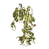
-Related structure data
| Related structure data |  6kbjC  6kbqSC  6kc2C  6li7C S: Starting model for refinement C: citing same article ( |
|---|---|
| Similar structure data |


-Links
 PDBj
PDBj- Assembly
| Deposited unit | 
| ||||||||
|---|---|---|---|---|---|---|---|---|---|
| 1 |
| ||||||||
| Unit cell |
|
-Components
-Protein , 1 types, 1 molecules A
| #1: Protein | / Lectin 1 Mass: 39004.953 Da / Num. of mol.: 1 / Source method: isolated from a natural source / Source: (natural) Pleurotus ostreatus (oyster mushroom) / References: UniProt: E7E2M2 |
|---|
-Sugars , 2 types, 4 molecules 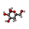
| #2: Polysaccharide | / Mass: 424.401 Da / Num. of mol.: 3 Source method: isolated from a genetically manipulated source #5: Sugar | ChemComp-GLA / | Galactose Type: D-saccharide, alpha linking / Mass: 180.156 Da / Num. of mol.: 1 Type: D-saccharide, alpha linking / Mass: 180.156 Da / Num. of mol.: 1Source method: isolated from a genetically manipulated source Formula: C6H12O6 / Feature type: SUBJECT OF INVESTIGATION |
|---|
-Non-polymers , 3 types, 125 molecules 


| #3: Chemical |  Mass: 40.078 Da / Num. of mol.: 2 / Source method: obtained synthetically / Formula: Ca Mass: 40.078 Da / Num. of mol.: 2 / Source method: obtained synthetically / Formula: Ca#4: Chemical | Glycerol Mass: 92.094 Da / Num. of mol.: 2 / Source method: obtained synthetically / Formula: C3H8O3 / Feature type: SUBJECT OF INVESTIGATION Mass: 92.094 Da / Num. of mol.: 2 / Source method: obtained synthetically / Formula: C3H8O3 / Feature type: SUBJECT OF INVESTIGATION#6: Water | ChemComp-HOH / | WaterMass: 18.015 Da / Num. of mol.: 121 / Source method: isolated from a natural source / Formula: H2O |
|---|
-Details
| Has ligand of interest | Y |
|---|
-Experimental details
-Experiment
| Experiment | Method: X-RAY DIFFRACTION / Number of used crystals: 1 |
|---|
- Sample preparation
| Crystal | Density Matthews: 6.29 Å3/Da / Density % sol: 80.43 % |
|---|---|
| Crystal grow | Temperature: 300 K / Method: vapor diffusion, hanging drop / pH: 5.5 / Details: 0.9M Na2HPO4/K2HPO4, pH 5.5, 33% glycerol |
-Data collection
| Diffraction | Mean temperature: 100 K / Serial crystal experiment: N |
|---|---|
| Diffraction source | Source: SYNCHROTRON / Site: APS  / Beamline: 22-ID / Wavelength: 0.99 Å / Beamline: 22-ID / Wavelength: 0.99 Å |
| Detector | Type: DECTRIS EIGER2 XE 16M / Detector: PIXEL / Date: Mar 1, 2018 / Details: Si 111 |
| Radiation | Monochromator: Rosenbaum-Rock double-crystal monochromator / Protocol: SINGLE WAVELENGTH / Monochromatic (M) / Laue (L): M / Scattering type: x-ray |
| Radiation wavelength | Wavelength: 0.99 Å / Relative weight: 1 |
| Reflection | Resolution: 2.4→30.04 Å / Num. obs: 39208 / % possible obs: 98.97 % / Redundancy: 5.1 % / CC1/2: 0.994 / CC star: 0.999 / Rpim(I) all: 0.043 / Rrim(I) all: 0.146 / Net I/σ(I): 12.98 |
| Reflection shell | Resolution: 2.4→2.49 Å / Mean I/σ(I) obs: 1.58 / Num. unique obs: 3803 / CC1/2: 0.604 / CC star: 0.868 / Rpim(I) all: 0.423 |
-Phasing
| Phasing | Method: molecular replacement |
|---|
- Processing
| Software |
| ||||||||||||||||||||||||||||||||||||||||||||||||||||||||||||
|---|---|---|---|---|---|---|---|---|---|---|---|---|---|---|---|---|---|---|---|---|---|---|---|---|---|---|---|---|---|---|---|---|---|---|---|---|---|---|---|---|---|---|---|---|---|---|---|---|---|---|---|---|---|---|---|---|---|---|---|---|---|
| Refinement | Method to determine structure: MOLECULAR REPLACEMENT Starting model: 6kbq Resolution: 2.4→30.04 Å / Cor.coef. Fo:Fc: 0.953 / Cor.coef. Fo:Fc free: 0.937 / SU B: 4.729 / SU ML: 0.104 / Cross valid method: THROUGHOUT / σ(F): 0 / ESU R: 0.146 / ESU R Free: 0.142 Details: HYDROGENS HAVE BEEN ADDED IN THE RIDING POSITIONS U VALUES : REFINED INDIVIDUALLY
| ||||||||||||||||||||||||||||||||||||||||||||||||||||||||||||
| Solvent computation | Ion probe radii: 0.8 Å / Shrinkage radii: 0.8 Å / VDW probe radii: 1.2 Å | ||||||||||||||||||||||||||||||||||||||||||||||||||||||||||||
| Displacement parameters | Biso max: 123.74 Å2 / Biso mean: 36.867 Å2 / Biso min: 20.72 Å2
| ||||||||||||||||||||||||||||||||||||||||||||||||||||||||||||
| Refinement step | Cycle: final / Resolution: 2.4→30.04 Å
| ||||||||||||||||||||||||||||||||||||||||||||||||||||||||||||
| Refine LS restraints |
| ||||||||||||||||||||||||||||||||||||||||||||||||||||||||||||
| LS refinement shell | Resolution: 2.402→2.465 Å / Rfactor Rfree error: 0 / Total num. of bins used: 20
|
