 Movie
Movie Controller


+ Open data

- Basic information
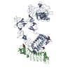
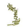
| Entry | Database: PDB / ID: 6lgx | ||||||
|---|---|---|---|---|---|---|---|
| Title | Structure of Rabies virus glycoprotein at basic pH | ||||||
 Components Components | Glycoprotein,Glycoprotein,Glycoprotein | ||||||
 Keywords Keywords |  VIRAL PROTEIN / functional class VIRAL PROTEIN / functional class | ||||||
| Function / homology | Rhabdovirus glycoprotein / Rhabdovirus spike glycoprotein / viral envelope / virion membrane / membrane / Glycoprotein / Glycoprotein / Glycoprotein / Glycoprotein Function and homology information Function and homology information | ||||||
| Biological species |  Rabies lyssavirus Rabies lyssavirus | ||||||
| Method | X-RAY DIFFRACTION / SYNCHROTRON / MOLECULAR REPLACEMENT / Resolution: 3.097 Å | ||||||
 Authors Authors | Yang, F.L. / Lin, S. / Ye, F. / Yang, J. / Qi, J.X. / Chen, Z.J. / Lin, X. / Wang, J.C. / Yue, D. / Cheng, Y.W. ...Yang, F.L. / Lin, S. / Ye, F. / Yang, J. / Qi, J.X. / Chen, Z.J. / Lin, X. / Wang, J.C. / Yue, D. / Cheng, Y.W. / Chen, Z.M. / Chen, H. / You, Y. / Zhang, Z.L. / Yang, Y. / Yang, M. / Sun, H.L. / Li, Y.H. / Cao, Y. / Yang, S.Y. / Wei, Y.Q. / Gao, G.F. / Lu, G.W. | ||||||
 Citation Citation | Journal: Cell Host Microbe / Year: 2020 Title: Structural Analysis of Rabies Virus Glycoprotein Reveals pH-Dependent Conformational Changes and Interactions with a Neutralizing Antibody. Authors: Yang, F. / Lin, S. / Ye, F. / Yang, J. / Qi, J. / Chen, Z. / Lin, X. / Wang, J. / Yue, D. / Cheng, Y. / Chen, Z. / Chen, H. / You, Y. / Zhang, Z. / Yang, Y. / Yang, M. / Sun, H. / Li, Y. / ...Authors: Yang, F. / Lin, S. / Ye, F. / Yang, J. / Qi, J. / Chen, Z. / Lin, X. / Wang, J. / Yue, D. / Cheng, Y. / Chen, Z. / Chen, H. / You, Y. / Zhang, Z. / Yang, Y. / Yang, M. / Sun, H. / Li, Y. / Cao, Y. / Yang, S. / Wei, Y. / Gao, G.F. / Lu, G. | ||||||
| History |
|
- Structure visualization
| Structure viewer | Molecule: MolmilJmol/JSmol |
|---|
- Downloads & links
-Download
| PDBx/mmCIF format | 6lgx.cif.gz | 300.7 KB | Display | PDBx/mmCIF format |
|---|---|---|---|---|
| PDB format | pdb6lgx.ent.gz | 245.4 KB | Display | PDB format |
| PDBx/mmJSON format | 6lgx.json.gz | Tree view | PDBx/mmJSON format | |
| Others |  Other downloads Other downloads |
-Validation report
| Arichive directory | https://data.pdbj.org/pub/pdb/validation_reports/lg/6lgxftp://data.pdbj.org/pub/pdb/validation_reports/lg/6lgx | HTTPS FTP |
|---|
-Related structure data
| Related structure data |  6lgwSC S: Starting model for refinement C: citing same article ( |
|---|---|
| Similar structure data |


-Links
 PDBj
PDBj- Assembly
| Deposited unit | 
| ||||||||
|---|---|---|---|---|---|---|---|---|---|
| 1 | 
| ||||||||
| Unit cell |
|
-Components
| #1: Protein | Mass: 49633.281 Da / Num. of mol.: 2 Source method: isolated from a genetically manipulated source Source: (gene. exp.) Rabies lyssavirus / Production host:  Trichoplusia ni (cabbage looper) Trichoplusia ni (cabbage looper)References: UniProt: Q5JZZ2, UniProt: Q2Z2I1, UniProt: D8VEC1, UniProt: O92284*PLUS |
|---|
-Experimental details
-Experiment
| Experiment | Method: X-RAY DIFFRACTION / Number of used crystals: 1 |
|---|
- Sample preparation
| Crystal | Density Matthews: 4.06 Å3/Da / Density % sol: 69.7 % |
|---|---|
| Crystal grow | Temperature: 291 K / Method: vapor diffusion, sitting drop / pH: 8 / Details: 0.1 M BICINE, 14% Polyethylene glycol 300 |
-Data collection
| Diffraction | Mean temperature: 100 K / Serial crystal experiment: N |
|---|---|
| Diffraction source | Source: SYNCHROTRON / Site: SSRF  / Beamline: BL17U1 / Wavelength: 0.97853 Å / Beamline: BL17U1 / Wavelength: 0.97853 Å |
| Detector | Type: DECTRIS PILATUS3 S 6M / Detector: PIXEL / Date: Nov 8, 2017 |
| Radiation | Protocol: SINGLE WAVELENGTH / Monochromatic (M) / Laue (L): M / Scattering type: x-ray |
| Radiation wavelength | Wavelength: 0.97853 Å / Relative weight: 1 |
| Reflection | Resolution: 3.097→50 Å / Num. obs: 28680 / % possible obs: 99.7 % / Observed criterion σ(F): 0 / Observed criterion σ(I): -3 / Redundancy: 6.8 % / Biso Wilson estimate: 106.98 Å2 / CC1/2: 0.985 / Net I/σ(I): 14.45 |
| Reflection shell | Resolution: 3.097→3.21 Å / Redundancy: 6.7 % / Num. unique obs: 2852 / CC1/2: 0.659 / % possible all: 99.7 |
- Processing
| Software |
| ||||||||||||||||||||||||||||||||||||||||||||||||||||||||||||||||||
|---|---|---|---|---|---|---|---|---|---|---|---|---|---|---|---|---|---|---|---|---|---|---|---|---|---|---|---|---|---|---|---|---|---|---|---|---|---|---|---|---|---|---|---|---|---|---|---|---|---|---|---|---|---|---|---|---|---|---|---|---|---|---|---|---|---|---|---|
| Refinement | Method to determine structure: MOLECULAR REPLACEMENT Starting model: 6LGW Resolution: 3.097→47.398 Å / SU ML: 0.47 / Cross valid method: THROUGHOUT / σ(F): 1.35 / Phase error: 36.37
| ||||||||||||||||||||||||||||||||||||||||||||||||||||||||||||||||||
| Solvent computation | Shrinkage radii: 0.9 Å / VDW probe radii: 1.11 Å | ||||||||||||||||||||||||||||||||||||||||||||||||||||||||||||||||||
| Displacement parameters | Biso max: 248.86 Å2 / Biso mean: 123.7361 Å2 / Biso min: 50.24 Å2 | ||||||||||||||||||||||||||||||||||||||||||||||||||||||||||||||||||
| Refinement step | Cycle: final / Resolution: 3.097→47.398 Å
| ||||||||||||||||||||||||||||||||||||||||||||||||||||||||||||||||||
| Refine LS restraints |
| ||||||||||||||||||||||||||||||||||||||||||||||||||||||||||||||||||
| LS refinement shell | Refine-ID: X-RAY DIFFRACTION / Rfactor Rfree error: 0
| ||||||||||||||||||||||||||||||||||||||||||||||||||||||||||||||||||
| Refinement TLS params. | Method: refined / Origin x: 42.622 Å / Origin y: 23.2452 Å / Origin z: 14.9364 Å
| ||||||||||||||||||||||||||||||||||||||||||||||||||||||||||||||||||
| Refinement TLS group |
|
