 Movie
Movie Controller

[English] 日本語
 Yorodumi
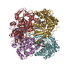
- PDB-6le4: Crystal structure of cystathionine gamma-lyase from Lactobacillus... -
+ Open data

- Basic information
| Entry | Database: PDB / ID: 6le4 | ||||||
|---|---|---|---|---|---|---|---|
| Title | Crystal structure of cystathionine gamma-lyase from Lactobacillus plantarum complexed with cystathionine | ||||||
 Components Components | Cystathionine gamma-lyase | ||||||
 Keywords Keywords | LYASE / cystathionine gamma-lyase | ||||||
| Function / homology |  Function and homology information Function and homology informationcystathionine gamma-synthase activity (acts on O-phosphohomoserine) / cystathionine gamma-synthase activity / cystathionine beta-lyase / cystathionine gamma-lyase / L-cystine L-cysteine-lyase (deaminating) / : / cystathionine gamma-lyase activity / L-cysteine desulfhydrase activity / transsulfuration / transaminase activity ...cystathionine gamma-synthase activity (acts on O-phosphohomoserine) / cystathionine gamma-synthase activity / cystathionine beta-lyase / cystathionine gamma-lyase / L-cystine L-cysteine-lyase (deaminating) / : / cystathionine gamma-lyase activity / L-cysteine desulfhydrase activity / transsulfuration / transaminase activity / pyridoxal phosphate binding / lyase activitySimilarity search - Function | ||||||
| Biological species |  Lactobacillus plantarum (bacteria) Lactobacillus plantarum (bacteria) | ||||||
| Method | X-RAY DIFFRACTION / SYNCHROTRON / MOLECULAR REPLACEMENT / Resolution: 3.1 Å | ||||||
 Authors Authors | Oda, K. / Matoba, Y. | ||||||
 Citation Citation | Journal: Sci Rep / Year: 2020 Title: Catalytic specificity of the Lactobacillus plantarum cystathionine gamma-lyase presumed by the crystallographic analysis. Authors: Matoba, Y. / Noda, M. / Yoshida, T. / Oda, K. / Ezumi, Y. / Yasutake, C. / Izuhara-Kihara, H. / Danshiitsoodol, N. / Kumagai, T. / Sugiyama, M. | ||||||
| History |
|
- Structure visualization
| Structure viewer | Molecule: MolmilJmol/JSmol |
|---|
- Downloads & links
-Download
| PDBx/mmCIF format | 6le4.cif.gz | 437.2 KB | Display | PDBx/mmCIF format |
|---|---|---|---|---|
| PDB format | pdb6le4.ent.gz | 360.1 KB | Display | PDB format |
| PDBx/mmJSON format | 6le4.json.gz | Tree view | PDBx/mmJSON format | |
| Others |  Other downloads Other downloads |
-Validation report
| Arichive directory | https://data.pdbj.org/pub/pdb/validation_reports/le/6le4ftp://data.pdbj.org/pub/pdb/validation_reports/le/6le4 | HTTPS FTP |
|---|
-Related structure data
| Related structure data |  6ldoC  4l0oS S: Starting model for refinement C: citing same article ( |
|---|---|
| Similar structure data |


-Links
 PDBj
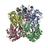
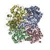
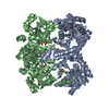
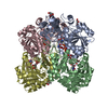
PDBj- Assembly
| Deposited unit | 
| ||||||||||||
|---|---|---|---|---|---|---|---|---|---|---|---|---|---|
| 1 | 
| ||||||||||||
| 2 | 
| ||||||||||||
| Unit cell |
| ||||||||||||
| Components on special symmetry positions |
|
-Components
| #1: Protein | / Cystathionine gamma-synthase Mass: 41915.562 Da / Num. of mol.: 6 / Fragment: Cystathionine gamma-lyase / Mutation: K194A Source method: isolated from a genetically manipulated source Source: (gene. exp.) Lactobacillus plantarum (bacteria) / Gene: BIZ32_00995, Nizo2891_3187 / Plasmid: pET21 / Production host: Escherichia coli BL21(DE3) (bacteria) / References: UniProt: A0A162EFJ4, UniProt: F9UT53*PLUS#2: Chemical | ChemComp-E9U / ( 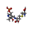  Mass: 451.389 Da / Num. of mol.: 6 / Source method: obtained synthetically / Formula: C15H22N3O9PS / Feature type: SUBJECT OF INVESTIGATION Mass: 451.389 Da / Num. of mol.: 6 / Source method: obtained synthetically / Formula: C15H22N3O9PS / Feature type: SUBJECT OF INVESTIGATION#3: Chemical | Phosphate  Mass: 94.971 Da / Num. of mol.: 3 / Source method: obtained synthetically / Formula: PO4 Mass: 94.971 Da / Num. of mol.: 3 / Source method: obtained synthetically / Formula: PO4#4: Water | ChemComp-HOH / | Water Mass: 18.015 Da / Num. of mol.: 296 / Source method: isolated from a natural source / Formula: H2O Mass: 18.015 Da / Num. of mol.: 296 / Source method: isolated from a natural source / Formula: H2OHas ligand of interest | Y | |
|---|
-Experimental details
-Experiment
| Experiment | Method: X-RAY DIFFRACTION / Number of used crystals: 1 |
|---|
- Sample preparation
| Crystal | Density Matthews: 4.42 Å3/Da / Density % sol: 72.14 % |
|---|---|
| Crystal grow | Temperature: 298 K / Method: vapor diffusion, sitting drop / pH: 4 / Details: KH2PO4, NaH2PO4 |
-Data collection
| Diffraction | Mean temperature: 100 K / Serial crystal experiment: N | |||||||||||||||||||||||||||||||||||||||||||||||||||||||||||||||||||||||||||||||||||||||||||||||||||
|---|---|---|---|---|---|---|---|---|---|---|---|---|---|---|---|---|---|---|---|---|---|---|---|---|---|---|---|---|---|---|---|---|---|---|---|---|---|---|---|---|---|---|---|---|---|---|---|---|---|---|---|---|---|---|---|---|---|---|---|---|---|---|---|---|---|---|---|---|---|---|---|---|---|---|---|---|---|---|---|---|---|---|---|---|---|---|---|---|---|---|---|---|---|---|---|---|---|---|---|---|
| Diffraction source | Source: SYNCHROTRON / Site: SPring-8  / Beamline: BL38B1 / Wavelength: 1 Å / Beamline: BL38B1 / Wavelength: 1 Å | |||||||||||||||||||||||||||||||||||||||||||||||||||||||||||||||||||||||||||||||||||||||||||||||||||
| Detector | Type: ADSC QUANTUM 315 / Detector: CCD / Date: May 30, 2014 / Details: mirros | |||||||||||||||||||||||||||||||||||||||||||||||||||||||||||||||||||||||||||||||||||||||||||||||||||
| Radiation | Monochromator: SINGLE WAVELENGTH / Protocol: SINGLE WAVELENGTH / Monochromatic (M) / Laue (L): M / Scattering type: x-ray | |||||||||||||||||||||||||||||||||||||||||||||||||||||||||||||||||||||||||||||||||||||||||||||||||||
| Radiation wavelength | Wavelength: 1 Å / Relative weight: 1 | |||||||||||||||||||||||||||||||||||||||||||||||||||||||||||||||||||||||||||||||||||||||||||||||||||
| Reflection | Resolution: 3.083→100 Å / Num. obs: 72733 / % possible obs: 91.4 % / Redundancy: 2 % / Rmerge(I) obs: 0.142 / Rpim(I) all: 0.123 / Rrim(I) all: 0.189 / Χ2: 1.01 / Net I/σ(I): 4.1 | |||||||||||||||||||||||||||||||||||||||||||||||||||||||||||||||||||||||||||||||||||||||||||||||||||
| Reflection shell | Diffraction-ID: 1
|
- Processing
| Software |
| ||||||||||||||||||||||||||||||||||||||||||||||||||||||||||||||||||||||||||||||||||||||||||||||||||||||||||||||||||||||||||||||||||||||||||||||||||||||||||||||||||
|---|---|---|---|---|---|---|---|---|---|---|---|---|---|---|---|---|---|---|---|---|---|---|---|---|---|---|---|---|---|---|---|---|---|---|---|---|---|---|---|---|---|---|---|---|---|---|---|---|---|---|---|---|---|---|---|---|---|---|---|---|---|---|---|---|---|---|---|---|---|---|---|---|---|---|---|---|---|---|---|---|---|---|---|---|---|---|---|---|---|---|---|---|---|---|---|---|---|---|---|---|---|---|---|---|---|---|---|---|---|---|---|---|---|---|---|---|---|---|---|---|---|---|---|---|---|---|---|---|---|---|---|---|---|---|---|---|---|---|---|---|---|---|---|---|---|---|---|---|---|---|---|---|---|---|---|---|---|---|---|---|---|---|---|
| Refinement | Method to determine structure: MOLECULAR REPLACEMENT Starting model: pdbid 4L0O Resolution: 3.1→36.257 Å / SU ML: 0.43 / Cross valid method: THROUGHOUT / σ(F): 1.34 / Phase error: 27.63
| ||||||||||||||||||||||||||||||||||||||||||||||||||||||||||||||||||||||||||||||||||||||||||||||||||||||||||||||||||||||||||||||||||||||||||||||||||||||||||||||||||
| Solvent computation | Shrinkage radii: 0.9 Å / VDW probe radii: 1.11 Å | ||||||||||||||||||||||||||||||||||||||||||||||||||||||||||||||||||||||||||||||||||||||||||||||||||||||||||||||||||||||||||||||||||||||||||||||||||||||||||||||||||
| Displacement parameters | Biso max: 341.95 Å2 / Biso mean: 39.3219 Å2 / Biso min: 0.77 Å2 | ||||||||||||||||||||||||||||||||||||||||||||||||||||||||||||||||||||||||||||||||||||||||||||||||||||||||||||||||||||||||||||||||||||||||||||||||||||||||||||||||||
| Refinement step | Cycle: final / Resolution: 3.1→36.257 Å
| ||||||||||||||||||||||||||||||||||||||||||||||||||||||||||||||||||||||||||||||||||||||||||||||||||||||||||||||||||||||||||||||||||||||||||||||||||||||||||||||||||
| LS refinement shell | Refine-ID: X-RAY DIFFRACTION / Rfactor Rfree error: 0
|
