 Movie
Movie Controller


+ Open data

- Basic information
| Entry | Database: PDB / ID: 6l5j | ||||||
|---|---|---|---|---|---|---|---|
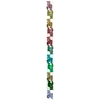
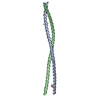
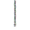
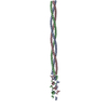
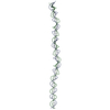
| Title | Crystal structure of human rootletin 1108-1317 | ||||||
 Components Components | Rootletin | ||||||
 Keywords Keywords | STRUCTURAL PROTEIN / centrosome / rootletin / coiled-coil | ||||||
| Function / homology |  Function and homology information Function and homology informationsubapical part of cell / establishment of organelle localization / ciliary basal body organization / centriole-centriole cohesion / 9+2 motile cilium / epithelial structure maintenance / protein localization to organelle / positive regulation of protein localization to cilium / positive regulation of cilium assembly / cellular homeostasis ...subapical part of cell / establishment of organelle localization / ciliary basal body organization / centriole-centriole cohesion / 9+2 motile cilium / epithelial structure maintenance / protein localization to organelle / positive regulation of protein localization to cilium / positive regulation of cilium assembly / cellular homeostasis / photoreceptor cell maintenance / centrosome cycle / ciliary rootlet / kinesin binding / photoreceptor inner segment / centriole / establishment of localization in cell / protein localization / structural constituent of cytoskeleton / actin cytoskeleton / actin binding / centrosome / structural molecule activity / extracellular exosome / plasma membrane / cytosolSimilarity search - Function | ||||||
| Biological species |  Homo sapiens (human) Homo sapiens (human) | ||||||
| Method | X-RAY DIFFRACTION / SYNCHROTRON / MAD / Resolution: 2.77 Å | ||||||
 Authors Authors | Kim, J. / Choi, H.J. | ||||||
| Funding support |  Korea, Republic Of, 1items Korea, Republic Of, 1items
| ||||||
 Citation Citation | Journal: J.Mol.Biol. / Year: 2020 Title: Identification of a Structurally Dynamic Domain for Oligomer Formation in Rootletin. Authors: Ko, D. / Kim, J. / Rhee, K. / Choi, H.J. | ||||||
| History |
|
- Structure visualization
| Structure viewer | Molecule: MolmilJmol/JSmol |
|---|
- Downloads & links
-Download
| PDBx/mmCIF format | 6l5j.cif.gz | 263.8 KB | Display | PDBx/mmCIF format |
|---|---|---|---|---|
| PDB format | pdb6l5j.ent.gz | 209 KB | Display | PDB format |
| PDBx/mmJSON format | 6l5j.json.gz | Tree view | PDBx/mmJSON format | |
| Others |  Other downloads Other downloads |
-Validation report
| Arichive directory | https://data.pdbj.org/pub/pdb/validation_reports/l5/6l5jftp://data.pdbj.org/pub/pdb/validation_reports/l5/6l5j | HTTPS FTP |
|---|
-Related structure data


-Links
 PDBj
PDBj- Assembly
| Deposited unit | 
| ||||||||
|---|---|---|---|---|---|---|---|---|---|
| 1 | 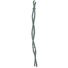
| ||||||||
| 2 | 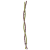
| ||||||||
| Unit cell |
|
-Components
| #1: Protein | / Ciliary rootlet coiled-coil protein Mass: 25118.439 Da / Num. of mol.: 4 / Mutation: L1153M, L1166M Source method: isolated from a genetically manipulated source Source: (gene. exp.) Homo sapiens (human) / Gene: CROCC, KIAA0445 / Production host:  Escherichia coli (E. coli) / References: UniProt: Q5TZA2 Escherichia coli (E. coli) / References: UniProt: Q5TZA2Has ligand of interest | N | |
|---|
-Experimental details
-Experiment
| Experiment | Method: X-RAY DIFFRACTION / Number of used crystals: 1 |
|---|
- Sample preparation
| Crystal | Density Matthews: 2.8 Å3/Da / Density % sol: 56.02 % |
|---|---|
| Crystal grow | Temperature: 278 K / Method: vapor diffusion, hanging drop / pH: 7.5 / Details: 0.1 M Tris pH 7.5, 4% PEG 6000 |
-Data collection
| Diffraction | Mean temperature: 100 K / Serial crystal experiment: N | |||||||||||||||||||||||||||||||||||||||||||||||||||||||||||||||||||||||||||||||||||||||||||||||||||||||||||||||||||||||||||||||||||||||||||||||||||||||||||||||||||||||||||||||||||||||||||||
|---|---|---|---|---|---|---|---|---|---|---|---|---|---|---|---|---|---|---|---|---|---|---|---|---|---|---|---|---|---|---|---|---|---|---|---|---|---|---|---|---|---|---|---|---|---|---|---|---|---|---|---|---|---|---|---|---|---|---|---|---|---|---|---|---|---|---|---|---|---|---|---|---|---|---|---|---|---|---|---|---|---|---|---|---|---|---|---|---|---|---|---|---|---|---|---|---|---|---|---|---|---|---|---|---|---|---|---|---|---|---|---|---|---|---|---|---|---|---|---|---|---|---|---|---|---|---|---|---|---|---|---|---|---|---|---|---|---|---|---|---|---|---|---|---|---|---|---|---|---|---|---|---|---|---|---|---|---|---|---|---|---|---|---|---|---|---|---|---|---|---|---|---|---|---|---|---|---|---|---|---|---|---|---|---|---|---|---|---|---|---|
| Diffraction source | Source: SYNCHROTRON / Site: PAL/PLS / Beamline: 5C (4A) / Wavelength: 0.97942 Å | |||||||||||||||||||||||||||||||||||||||||||||||||||||||||||||||||||||||||||||||||||||||||||||||||||||||||||||||||||||||||||||||||||||||||||||||||||||||||||||||||||||||||||||||||||||||||||||
| Detector | Type: DECTRIS PILATUS3 6M / Detector: PIXEL / Date: Mar 25, 2019 | |||||||||||||||||||||||||||||||||||||||||||||||||||||||||||||||||||||||||||||||||||||||||||||||||||||||||||||||||||||||||||||||||||||||||||||||||||||||||||||||||||||||||||||||||||||||||||||
| Radiation | Protocol: MAD / Monochromatic (M) / Laue (L): M / Scattering type: x-ray | |||||||||||||||||||||||||||||||||||||||||||||||||||||||||||||||||||||||||||||||||||||||||||||||||||||||||||||||||||||||||||||||||||||||||||||||||||||||||||||||||||||||||||||||||||||||||||||
| Radiation wavelength | Wavelength: 0.97942 Å / Relative weight: 1 | |||||||||||||||||||||||||||||||||||||||||||||||||||||||||||||||||||||||||||||||||||||||||||||||||||||||||||||||||||||||||||||||||||||||||||||||||||||||||||||||||||||||||||||||||||||||||||||
| Reflection | Resolution: 2.77→50 Å / Num. obs: 25990 / % possible obs: 95.1 % / Redundancy: 3.5 % / Rmerge(I) obs: 0.1 / Rpim(I) all: 0.064 / Rrim(I) all: 0.119 / Χ2: 1.246 / Net I/σ(I): 4.8 | |||||||||||||||||||||||||||||||||||||||||||||||||||||||||||||||||||||||||||||||||||||||||||||||||||||||||||||||||||||||||||||||||||||||||||||||||||||||||||||||||||||||||||||||||||||||||||||
| Reflection shell | Diffraction-ID: 1
|
- Processing
| Software |
| ||||||||||||||||||||||||||||||||||||||||||||||||||||||||||||
|---|---|---|---|---|---|---|---|---|---|---|---|---|---|---|---|---|---|---|---|---|---|---|---|---|---|---|---|---|---|---|---|---|---|---|---|---|---|---|---|---|---|---|---|---|---|---|---|---|---|---|---|---|---|---|---|---|---|---|---|---|---|
| Refinement | Method to determine structure: MAD / Resolution: 2.77→47.473 Å / SU ML: 0.32 / Cross valid method: THROUGHOUT / σ(F): 1.98 / Phase error: 41.96
| ||||||||||||||||||||||||||||||||||||||||||||||||||||||||||||
| Solvent computation | Shrinkage radii: 0.9 Å / VDW probe radii: 1.11 Å | ||||||||||||||||||||||||||||||||||||||||||||||||||||||||||||
| Displacement parameters | Biso max: 169.81 Å2 / Biso mean: 79.8283 Å2 / Biso min: 38.29 Å2 | ||||||||||||||||||||||||||||||||||||||||||||||||||||||||||||
| Refinement step | Cycle: final / Resolution: 2.77→47.473 Å
| ||||||||||||||||||||||||||||||||||||||||||||||||||||||||||||
| LS refinement shell | Refine-ID: X-RAY DIFFRACTION / Rfactor Rfree error: 0
|
